. 2012 Aug 21;109(34):13602-7.
doi: 10.1073/pnas.1204593109. Epub 2012 Aug 3.

Structure of the yeast F1Fo-ATP synthase dimer and its role in shaping the mitochondrial cristae

Affiliations

Structure of the yeast F1Fo-ATP synthase dimer and its role in shaping the mitochondrial cristae

Karen M Davies et al. Proc Natl Acad Sci U S A. .

Abstract

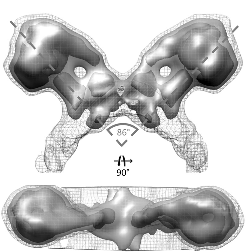
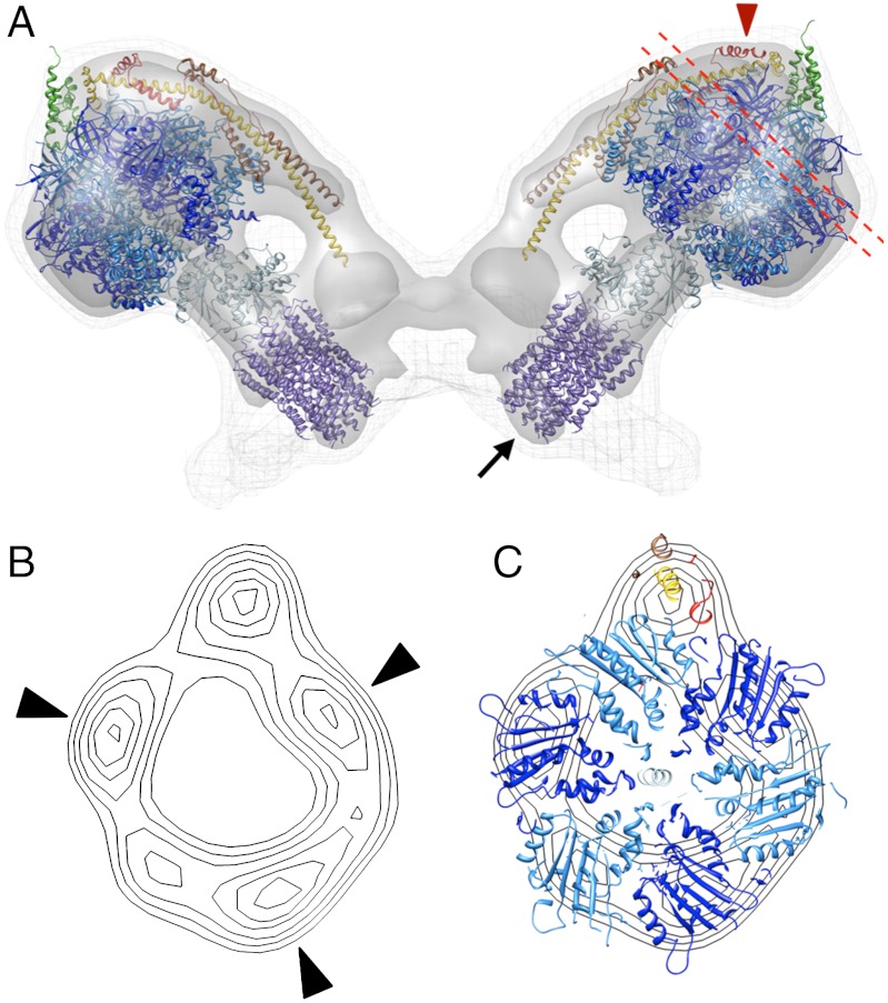
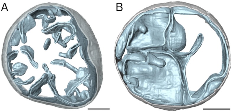
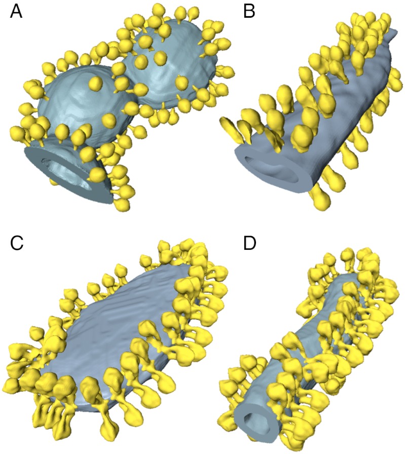
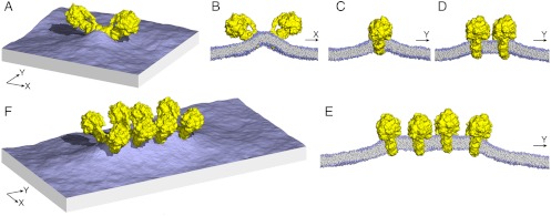
We used electron cryotomography of mitochondrial membranes from wild-type and mutant Saccharomyces cerevisiae to investigate the structure and organization of ATP synthase dimers in situ. Subtomogram averaging of the dimers to 3.7 nm resolution revealed a V-shaped structure of twofold symmetry, with an angle of 86° between monomers. The central and peripheral stalks are well resolved. The monomers interact within the membrane at the base of the peripheral stalks. In wild-type mitochondria ATP synthase dimers are found in rows along the highly curved cristae ridges, and appear to be crucial for membrane morphology. Strains deficient in the dimer-specific subunits e and g or the first transmembrane helix of subunit 4 lack both dimers and lamellar cristae. Instead, cristae are either absent or balloon-shaped, with ATP synthase monomers distributed randomly in the membrane. Computer simulations indicate that isolated dimers induce a plastic deformation in the lipid bilayer, which is partially relieved by their side-by-side association. We propose that the assembly of ATP synthase dimer rows is driven by the reduction in the membrane elastic energy, rather than by direct protein contacts, and that the dimer rows enable the formation of highly curved ridges in mitochondrial cristae.


Conflict of interest statement

The authors declare no conflict of interest.

Figures

Fig. 1.
Fig. 1.
Subtomogram average of the ATP synthase dimer from yeast mitochondria. Two ATP synthase monomers form a V-shaped dimer with an angle of 86° between their long axes. The central and peripheral stalks are clearly resolved. The dimer interface is located in the membrane between the two peripheral stalks. Threshold levels: mesh, 1σ; light grey, 2σ; dark grey, 3σ. See also Movie S1.
Fig. 2.
Fig. 2.
Fitted atomic models of ATP synthase dimer. (A) side view of subtomogram average 3D volume with fitted atomic models. Blue-purple, F1/rotor ring assembly [PDB: 2WPD (12)]; green, OSCP [PDB:2B05 (25)]; yellow-red, peripheral stalk fragment [PDB: 2CLY (14)] with additional residues from [PDB: 2WSS (15)]. Arrow indicates the N-terminal ends of the rotor ring, which account for the bulge protruding into the cristae space; red arrowhead indicates subunit h. (B) contour map of projected density slice between the dashed red lines in (A). (C) same as (B) but with fitted atomic models. The F1 density has near-sixfold symmetry with three stronger density peaks (black arrowheads) alternating with weaker peaks. The β subunits (deep blue) were positioned in the strong densities with the peripheral stalk complex adjacent to a non-catalytic interface. See Movie S1.
Fig. 3.
Fig. 3.
Morphology of mitochondria from wild-type and mutant yeast strains. Surface-rendered volume of a mitochondrion from (A) wild-type and (B) the su4ΔTM1 yeast strain. Wild-type mitochondria have lamellar cristae with highly curved edges whereas mitochondria from mutants lacking either subunit e, g, or su4TM1 contain a number of separate inner membrane vesicles but few or no cristae. When cristae were present, they tended to be balloon-shaped with smooth, gently curving surfaces. Light grey-outer membrane, sky blue-inner membrane. (Scale bar, 200 nm). See also Fig. S4.
Fig. 4.
Fig. 4.
ATP synthase distribution in isolated mitochondrial membranes. Surface-rendered volumes of mitochondrial membranes from yeast strains lacking subunit g (A, B), and wild type (C, D). In the mutants, the ATP synthase complexes are monomeric, and randomly distributed over flat or gently curving membrane regions (A, B). By contrast ATP synthase from wild-type mitochondria form rows of dimers along the highly curved ridges of tubular (D) or disk-shaped (C) cristae vesicles.
Fig. 5.
Fig. 5.
Membrane curvature induced by ATP synthase dimers. (A) perspective view of a simulated membrane patch with an ATP synthase dimer distorting the planar lipid bilayer. The simulation is based on a coarse-grained representation of the dimer structure and its environment (see Methods); for clarity, the solvent is omitted. (B, C) cross sections through the membrane patch in (A) showing the curvature profile of the lipid bilayer in x and y direction. (D) and (E), curvature profiles as in (C) for membranes with two or four ATP synthase dimers, side by side. Note how the membrane deformation in y direction is relieved when two or more dimers assemble into a row. (F) perspective view of the row of four dimers shown in (E). 2D curvature maps are compared quantitatively in Fig. S6.

References

    1. Kagawa Y, Racker E. Partial resolution of the enzymes catalyzing oxidative phosphorylation. X. Correlation of morphology and function in submitochondrial particles. J Biol Chem. 1966;241:2475–2482. - PubMed
    1. Gilkerson RW, Selker JM, Capaldi RA. The cristal membrane of mitochondria is the principal site of oxidative phosphorylation. FEBS Lett. 2003;546:355–358. - PubMed
    1. Arnold I, Pfeiffer K, Neupert W, Stuart RA, Schagger H. Yeast mitochondrial F1F0-ATP synthase exists as a dimer: identification of three dimer-specific subunits. EMBO J. 1998;17:7170–7178. - PMC - PubMed
    1. Allen R, Schroeder C, Fok A. An investigation of mitochondrial inner membranes by rapid-freeze deep-etch techniques. J Cell Biol. 1989;108:2233–2240. - PMC - PubMed
    1. Davies KM, et al. Macromolecular organization of ATP synthase and complex I in whole mitochondria. Proc Natl Acad Sci USA. 2011;108:14121–14126. - PMC - PubMed

Publication types

Associated data