. 2012 Nov;350(2):199-213.
doi: 10.1007/s00441-012-1468-7. Epub 2012 Aug 3.

Interstitial cells in the primate gastrointestinal tract

Affiliations

Interstitial cells in the primate gastrointestinal tract

Peter J Blair et al. Cell Tissue Res. 2012 Nov.

Abstract

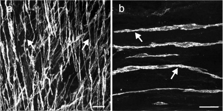
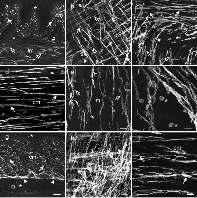
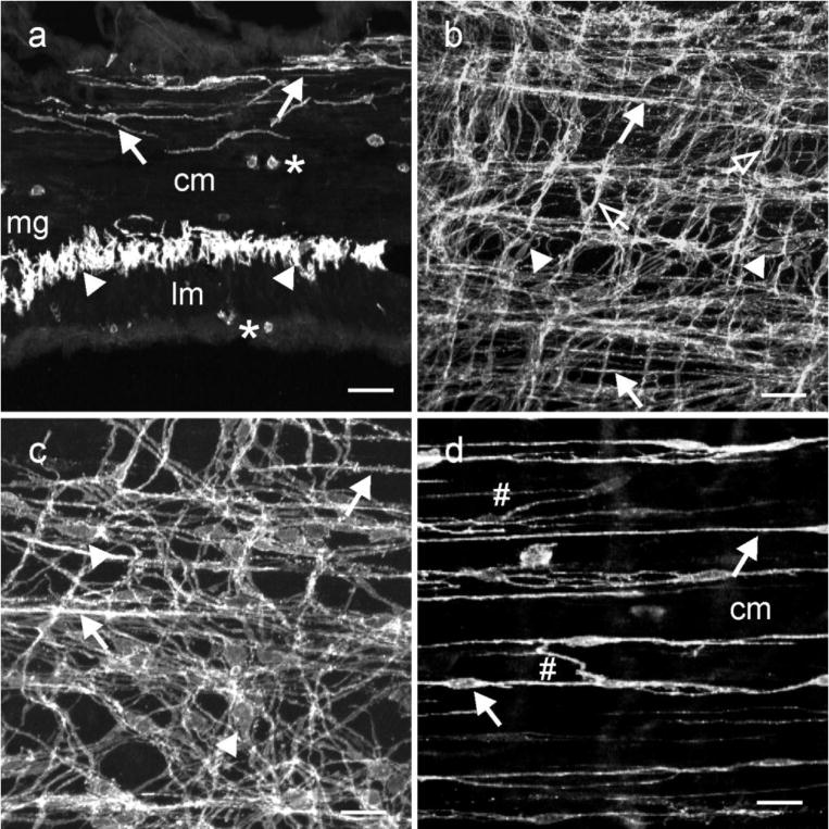
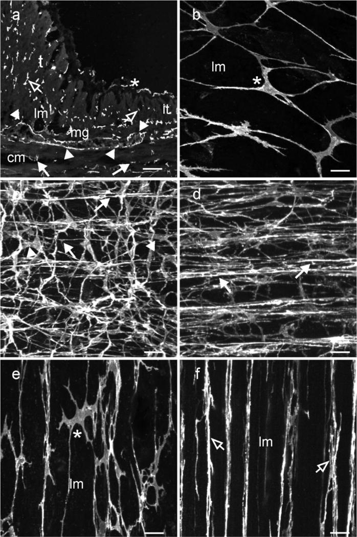
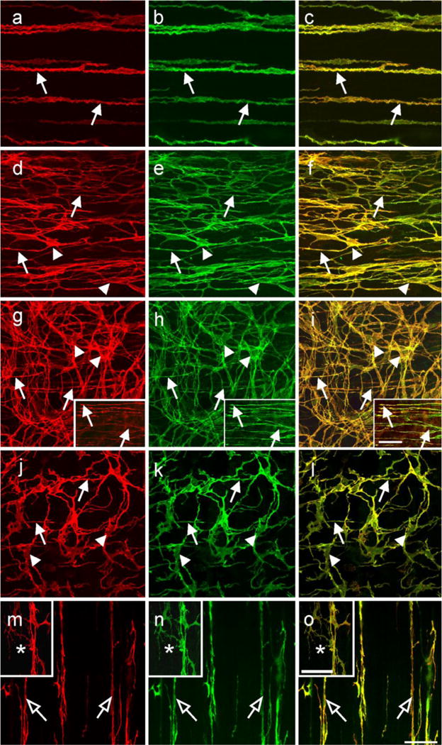
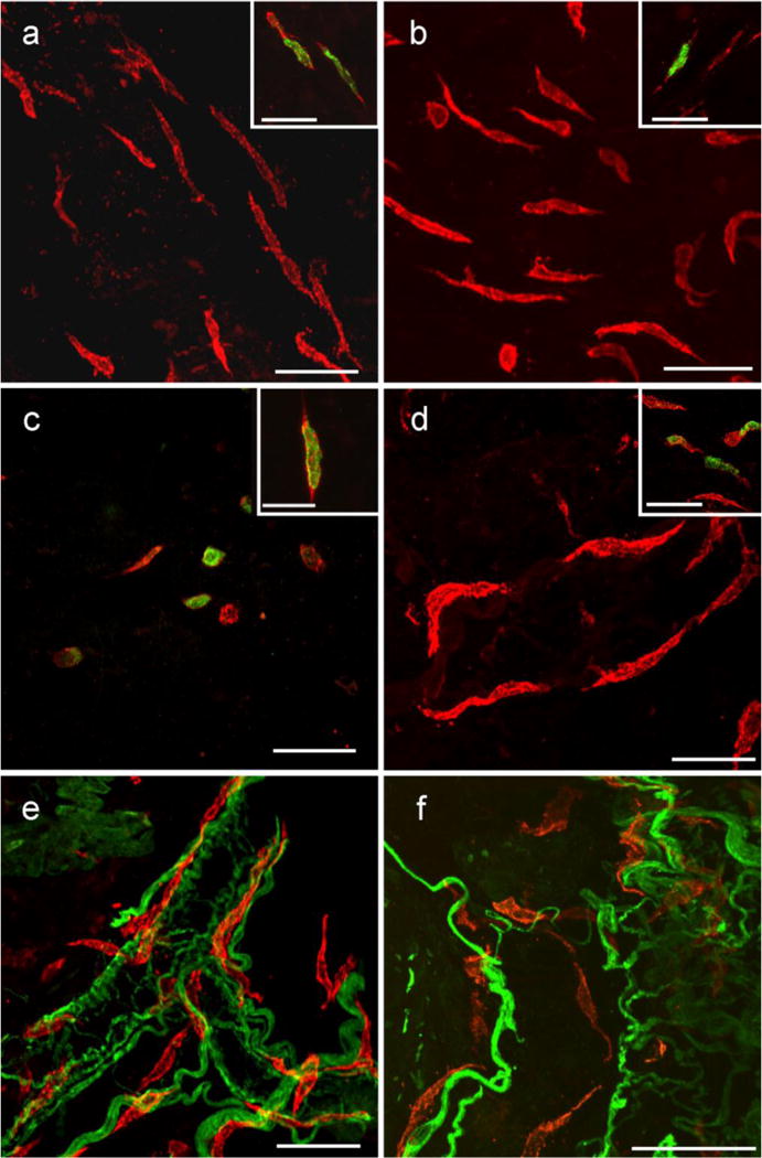
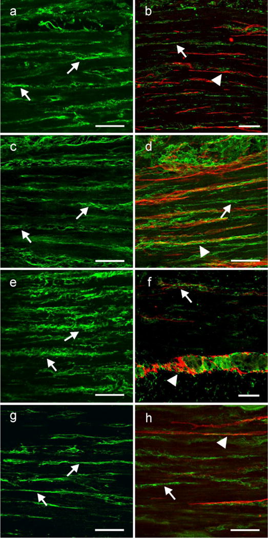
Kit immunohistochemistry and confocal reconstructions have provided detailed 3-dimensional images of ICC networks throughout the gastrointestinal (GI) tract. Morphological criteria have been used to establish that different classes of ICC exist within the GI tract and physiological studies have shown that these classes have distinct physiological roles in GI motility. Structural studies have focused predominately on rodent models and less information is available on whether similar classes of ICC exist within the GI tracts of humans or non-human primates. Using Kit immunohistochemistry and confocal imaging, we examined the 3-dimensional structure of ICC throughout the GI tract of cynomolgus monkeys. Whole or flat mounts and cryostat sections were used to examine ICC networks in the lower esophageal sphincter (LES), stomach, small intestine and colon. Anti-histamine antibodies were used to distinguish ICC from mast cells in the lamina propria. Kit labeling identified complex networks of ICC populations throughout the non-human primate GI tract that have structural characteristics similar to that described for ICC populations in rodent models. ICC-MY formed anastomosing networks in the myenteric plexus region. ICC-IM were interposed between smooth muscle cells in the stomach and colon and were concentrated within the deep muscular plexus (ICC-DMP) of the intestine. ICC-SEP were found in septal regions of the antrum that separated circular muscle bundles. Spindle-shaped histamine(+) mast cells were found in the lamina propria throughout the GI tract. Since similar sub-populations of ICC exist within the GI tract of primates and rodents and the use of rodents to study the functional roles of different classes of ICC is warranted.


Figures

Fig. 1
Fig. 1
ICC in the lower esophageal sphincter. (a, b) digital reconstructions of confocal images from flat cryosections (100 μm) of the LES. Kit+ ICC (arrows) possessed spindle shaped morphology with a central oval nucleus and were interspersed within the circular and longitudinal muscle layers. The dense population of ICC-IM ran parallel to the longitudinal axis of the muscle fibers. Scale bars = 50 μm in (a) and 25 μm in (b)
Fig. 2
Fig. 2
Distribution of ICC in the stomach. (a–f) ICC within the gastric fundus. (a) is a cryostat cross section through the fundus wall and (b) a whole mount preparation of the fundus tunica muscularis. Spindle shaped Kit+ ICC were dispersed within the circular (cm, solid arrows) and longitudinal muscle layers (lm, open arrows). The long axis of the ICC-IM cell bodies ran parallel to the smooth muscle cells of the respective muscle layers. (a,c) A distinct population of ICC was not observed adjacent to the myenteric border but ICC-IM from both muscle layers interconnected with each other and transversed both muscle layers (#). (b) Dense aggregations of ICC-IM in the circular layer ($) appeared to form networks within septa and around muscle bundles and may be analogous to septal ICC (ICC-SEP). (d–e) Not all ICC-IM in the fundus were spindle shaped (d) but cells in the longitudinal layer adjacent to the myenteric region (e) possessed several processes that often contacted adjacent ICC-IM. (f) At higher power, ICC-IM in both layers displayed spiny protrusions extending from the bi-lateral processes. Numerous rounded mast cells (*) were observed throughout the gastric fundus. (g–i) ICC within the gastric antrum. (g) is a cryosection cut transverse to the circular layer showing ICC-IM in the circular (cm, arrows) but not the longitudinal (lm) layer. ICC-SEP were observed to surround muscle bundles ($). A dense network of ICC-MY were observed interspersed between and surrounding myenteric ganglia (arrowheads), that can be seen in a flat mount section (h). ICC-MY possessed several processes that extended from a central nuclear region and contacted adjacent ICC-MY to form a distinct network (arrowheads). (i) Spindle shaped ICC-IM (arrows) ran parallel to the long axis of the circular layer (cm). ICC-IM possessed lateral processes that extended from the main body and contacted adjacent ICC-IM forming a loose network. Aggregations of ICC-IM formed rope-like networks, reminiscent of ICC-SEP ($), within the circular layer. Numerous mast cells were observed in the longitudinal layer and along the submucosal surface of the circular layer (*). Scale bars = 100 μm in (a & g), 50 μm in (b,c,d,e,h) and 25 μm in (f & h)
Fig. 3
Fig. 3
ICC within the small intestine. (a) is a cryostat section cut parallel to the circular layer. ICC-IM and ICC-DMP ran parallel to the long axis of smooth muscle fibers within the circular layer (cm, solid arrows). ICC-MY (arrowheads) can be observed within the intermuscular plane between the circular and longitudinal muscle layers (lm) at the level of the myenteric plexus (mg) at higher power. Occasional mast cells (*) were observed along the serosal aspect of the lm and within the cm. (b,c) confocal reconstructions of whole mounts through the jejunum at different magnifications. A dense anastomosing network of ICC-MY was observed at the level of the myenteric plexus that formed connections with adjacent ICC-MY (arrowheads). ICC-IM were also observed along the inner aspect of the circular layer (closed arrows) and occasionally in the longitudinal layer (open arrows). (d) is a digital reconstruction of ICC-IM through the circular layer. ICC-DMP were observed on the inner aspect of the circular layer (arrows) and ICC-IM were observed within the circular layer. ICC-DMP and ICC-IM possessed lateral projections that formed a loose interconnecting network with other ICC-IM (#;d). Scale bars = 50 μm in (d & b) and 25 μm in (c & d)
Fig. 4
Fig. 4
Distribution of ICC within the proximal colon. Kit+ ICC were distributed at several anatomical locations within the colonic wall and their density differed in taenia versus inter taenia regions. (a) shows a cryostat section through a region of the colonic wall containing inter taenia (it) and a more pronounced taenia (t) muscle. ICC-MY (arrowheads) can be seen surrounding a myenteric ganglia (mg), located between the circular (cm) and longitudinal muscle (lm) layers. ICC-IM are present within the circular (solid arrows) and longitudinal muscle layers (open arrows). (b–d) show whole mounts of Kit+ ICC along the serosal surface (a,b,e; *) of an inter-taenia region forming a loose interconnecting network and ICC-MY at the level of the myenteric plexus forming a dense anastomosing network (arrowheads; c). The processes that extended from a triangular or oval perinuclear region often bifurcated into multiple processes and contacted adjacent ICC-MY. (d) Shows a dense population of ICC-IM within the circular muscle layer. ICC-IM appeared to form rope-like networks, processes extended from the main axis of ICC-IM to connect with adjacent ICC-IM. ICC-IM were also observed within the longitudinal muscle layer where they ran parallel to smooth muscle cells. (e,f) reveal several distinct populations of ICC within the taenia. Panel (e) shows ICC-SS in the taenia that formed a loose interconnecting network (*). The main axis of these cells ran parallel with the longitudinal muscle cells and lateral projections contacted adjacent ICC forming an interconnecting network. (f) shows a second population of ICC within taenia. This population of ICC-IM was spindle shaped, were distributed throughout the band of muscle and ran parallel to the smooth muscle cells. ICC-IM did not possess the numerous lateral projections seen in ICC along the serosal surface. Scale bars = 100 μm in (a) and 25 μm in (b,c,d,e & f)
Fig. 5
Fig. 5
Cellular co-localization of Kit and ANO1/TMEM16A in ICC throughout the monkey GI tract. (a–c) show Kit (a; red) and TMEM16A (b; green) in ICC-IM (arrows) of the gastric fundus circular muscle. (c) shows a digital overlay of panels (a,b). Both Kit and ANO1 were co-localized in ICC-IM (arrows, yellow). (d–f) show Kit (d; red) and ANO1 (e; green) in ICC-IM (arrows) and ICC-MY (arrow heads) within the gastric antrum. Both Kit and ANO1 were co-localized within both populations of ICC (f; yellow). (g–i) show Kit (g; red) and ANO1 (h; green) in ICC at the level of the deep muscular plexus (ICC-DMP; arrows) and ICC-MY (arrowheads) of the small intestine. Digital overlay of (g,h) is shown in (i). Kit and ANO1 were co-localized in both ICC-DMP and ICC-MY (i; yellow) in the small intestine. Digital images of Kit and ANO1 labeling within ICC-DMP and their co-localization are shown in the insets of panels (g–i) (arrows). (j,k) show Kit (j; red) and ANO1 (k; green) labeling in ICC-IM (arrows) and ICC-MY (arrowheads) within the proximal colon. Co-localization of Kit and ANO1 is shown in (l; yellow). Panels (m–o) show Kit and ANO1-LI cellular co-localization in different populations of ICC of the taenia. Intramuscular ICC (open arrows) were both Kit (m; red) and ANO1 (n; green) immunopositive (as shown in the digital overlay in panel o). Serosal ICC (*) in the taenia were also Kit and ANO1+ (m–o, insets). Scale bar in (o) = 50 μm and applies to all panels. Scale bar in the inset in (i) = 50 μm and applies to insets in (g–i). Scale bar in inset in (o) = 50 μm and applies to insets in (m–o)
Fig. 6
Fig. 6
Kit+ cells within the submucosa and lamina propria of the GI tract. Flat sections and whole mount preparations of the mucosa and underlying submucosa from stomach, small intestine and colon. Kit+ cells within the submucosa and lamina propria in the gastric fundus (a), antrum (b), small intestine (c) and colon (d) were spindle shaped and did not appear to have a distinct orientation except when in close association with submucosal blood vessels (e,f colon shown). In the small intestine the number of these Kit+ cells that were spindle shaped was less than in other organs, but a larger number of rounded Kit+ cells were observed in the intestinal submucosa. Double labeling with PGP 9.5 and Kit revealed that the spindle shaped cells ran close to but were not intimately associated with nerves. PGP 9.5 labeling also identified autonomic nerves associated with submucosal blood vessels and spindle shaped cells were closely associated with these vessels (e). Double labeling with Kit and histamine revealed a sub-population of these spindle shaped cells were histamine+ in the stomach, small intestine and colon, suggesting these cells were likely mast cells (insets in a–d). Scale bar in (o) = 50 μm and applies to all panels. Scale bar in the inset in (i) = 50 μm and applies to insets in (g–i). Scale bar in all panels and insets = 50 μm, except inset in (c) = 25 μm
Fig. 7
Fig. 7
PDGFRα is expressed in a separate population of interstitial cells that are not Kit+. (a,c,e,g) Shows the cellular distribution of PDGFRα within the circular muscle layer of the gastric fundus, antrum small intestine and colon, respectively. PDGFRα + cells (arrows) ran parallel to the long axis of the circular muscle fibers. (b,d,f h) show double labeling of PDGFRα (green; arrows) and Kit (red; arrow heads) in two populations of cells in the fundus, antrum, small intestine and colon, respectively. Although the two cell populations were closely apposed to one another they were distinct, providing evidence that PDGFRα+ cells were not Kit+ ICC. Scale bars in all panels = 50 μm

Similar articles

Cited by

References

    1. Albertí E, Mikkelsen HB, Wang XY, Díaz M, Larsen JO, Huizinga JD, Jiménez M. Pacemaker activity and inhibitory neurotransmission in the colon of Ws/Ws mutant rats. Am J Physiol Gastrointest Liver Physiol. 2007;292:G1499–1510. - PubMed
    1. Aranishi H, Kunisawa Y, Komuro T. Characterization of interstitial cells of Cajal in the subserosal layer of the guinea-pig colon. Cell Tissue Res. 2009;335:323–329. - PubMed
    1. Beckett EA, Horiguchi K, Khoyi M, Sanders KM, Ward SM. Loss of enteric motor neurotransmission in the gastric fundus of Sl/Sl(d) mice. J Physiol. 2002;543:871–887. - PMC - PubMed
    1. Bernardini N, Segnani C, Ippolito C, De Giorgio R, Colucci R, Faussone-Pellegrini MS, Chiarugi M, Campani D, Castagna M, Mattii L, Blandizzi C, Dolfi A. Immunohistochemical Analysis of Myenteric Ganglia and Interstitial Cells of Cajal in Ulcerative Colitis. J Cell Mol Med 2011 - PMC - PubMed
    1. Burns AJ, Herbert TM, Ward SM, Sanders KM. Interstitial cells of Cajal in the guinea-pig gastrointestinal tract as revealed by c-Kit immunohistochemistry. Cell Tissue Res. 1997;290:11–20. - PubMed

Publication types

LinkOut - more resources