. 2013 Jan;62(1):149-59.
doi: 10.1007/s00262-012-1326-1. Epub 2012 Aug 4.

Memory T cells are uniquely resistant to melanoma-induced suppression

Affiliations

Memory T cells are uniquely resistant to melanoma-induced suppression

Lucy Wentworth et al. Cancer Immunol Immunother. 2013 Jan.

Abstract

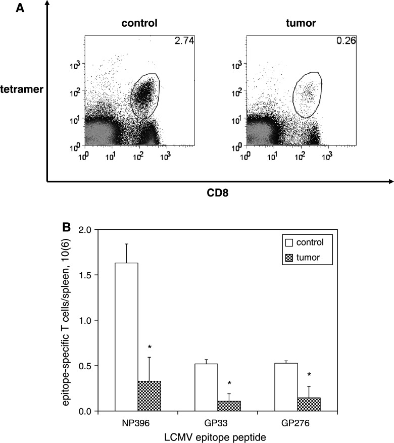
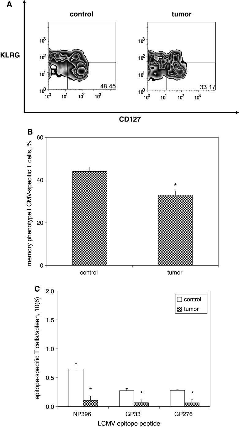
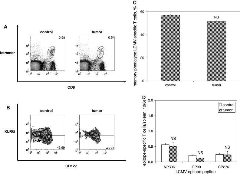
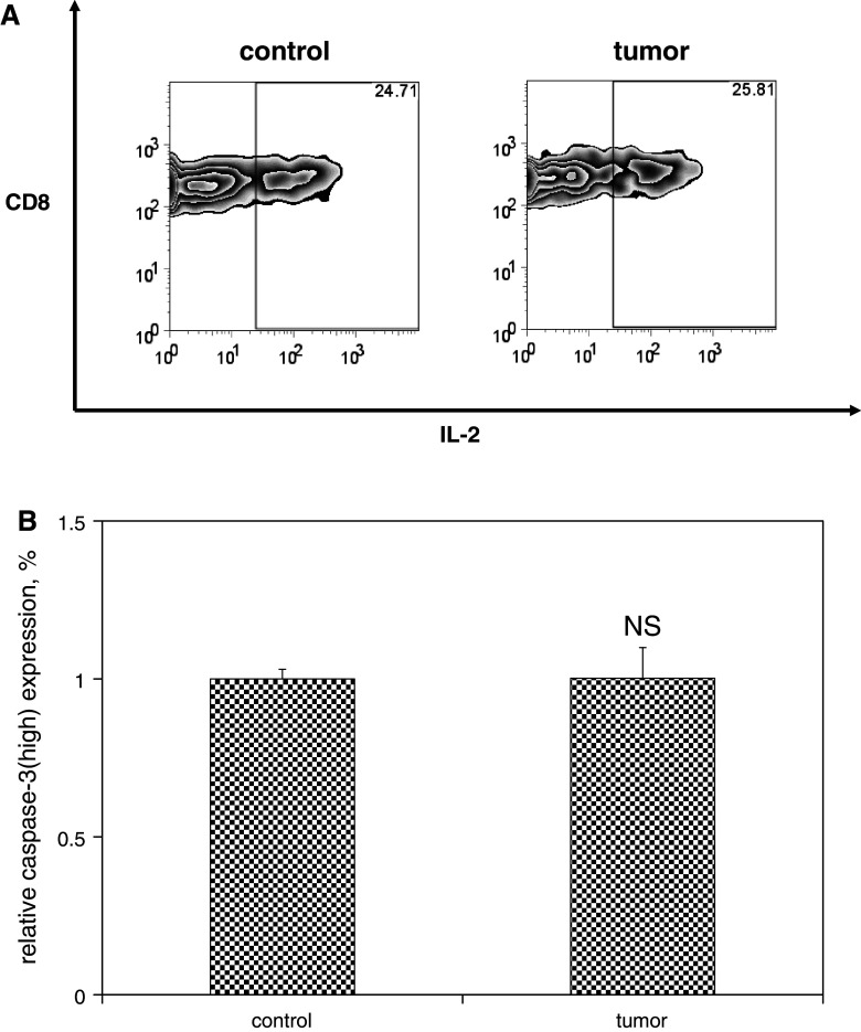
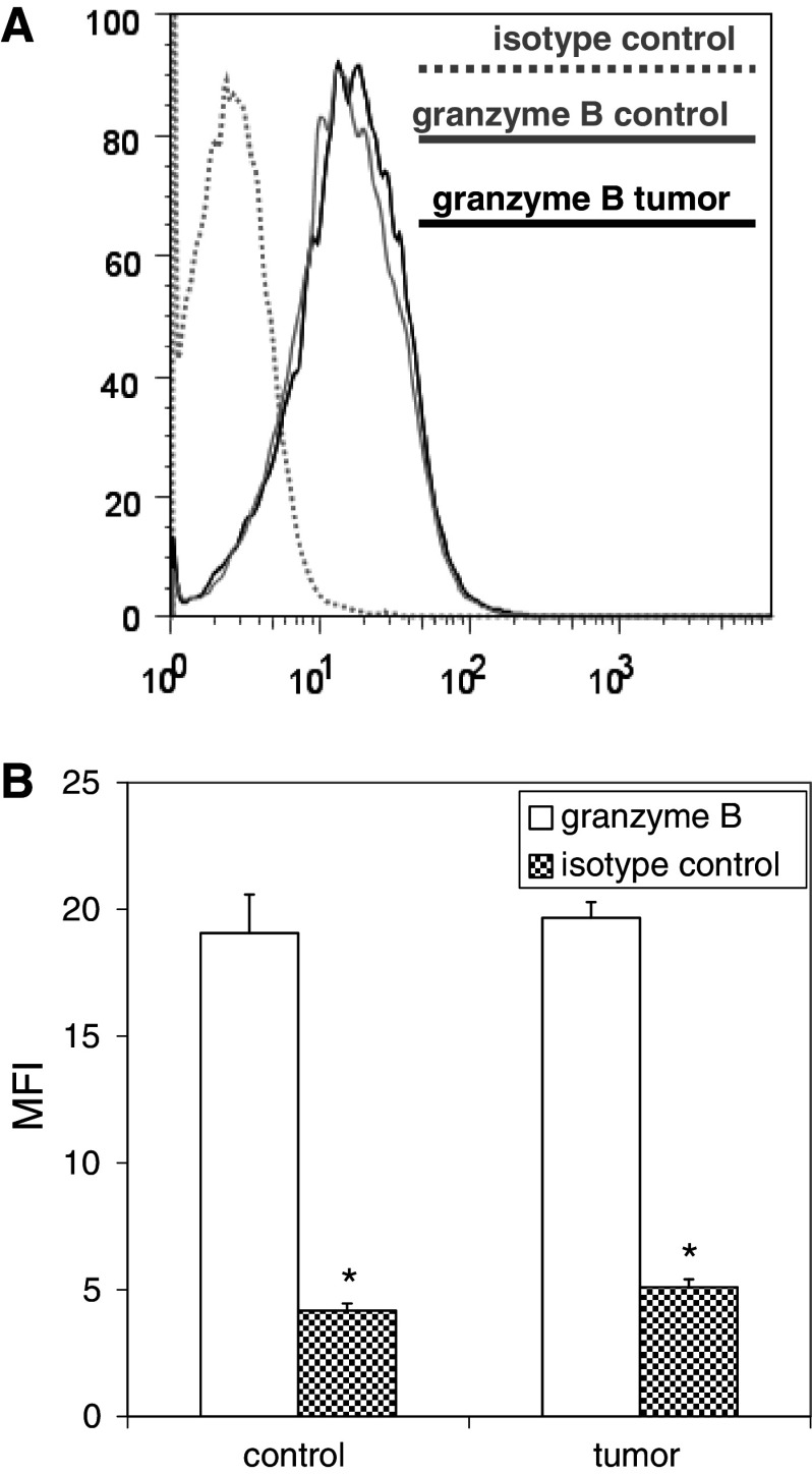
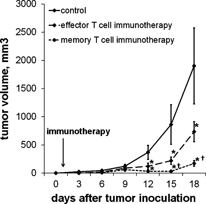
We have previously observed that in vivo exposure to growing melanoma tumors fundamentally alters activated T cell homeostasis by suppressing the ability of naïve T cells to undergo antigen-driven proliferative expansion. We hypothesized that exposure of T cells in later stages of differentiation to melanoma would have similar suppressive consequences. C57BL/6 mice were inoculated with media or syngeneic B16F10 melanoma tumors 8 or 60 days after infection with lymphocytic choriomeningitis virus (LCMV), and splenic populations of LCMV-specific T cells were quantified using flow cytometry 18 days after tumor inoculation. Inoculation with melanoma on post-infection day 8 potentiated the contraction of previously activated T cells. This enhanced contraction was associated with increased apoptotic susceptibility among T cells from tumor-bearing mice. In contrast, inoculation with melanoma on post-infection day 60 did not affect the ability of previously established memory T cells to maintain themselves in stable numbers. In addition, the ability of previously established memory T cells to respond to LCMV challenge was unaffected by melanoma. Following adoptive transfer into melanoma-bearing mice, tumor-specific memory T cells were significantly more effective at controlling melanoma growth than equivalent numbers of tumor-specific effector T cells. These observations suggest that memory T cells are uniquely resistant to suppressive influences exerted by melanoma on activated T cell homeostasis; these findings may have implications for T cell-based cancer immunotherapy.


Figures

Fig. 1
Fig. 1
Contraction of activated T cells is potentiated in the presence of melanoma. C57BL/6 mice were infected with LCMV then inoculated with media (n = 4) or B16F10 melanoma (n = 4) on post-infection day 8, at which time expansion of LCMV-specific CD8+ T cells reaches its peak and LCMV is cleared. Splenocytes were harvested 18 days later on post-infection day 26, at which time contraction of LCMV-specific T cells has occured. LCMV-specific CD8+ T cells were identified by staining cells with fluorophore-labeled antibodies against CD8 and fluorophore-labeled MHC tetramers loaded with MHC class I-restricted LCMV epitope peptides, and then quantified by flow cytometric analysis. a Representative dot plots demonstrate smaller percentages of CD8+ T cells specific for the immunodominant LCMV epitope peptide NP396 (gated on lymphocytes) among splenocytes harvested from melanoma-bearing mice compared with control mice. b Histograms of absolute numbers of LCMV-specific CD8+ T cells per spleen (calculated as the percentage of cells by flow cytometry multiplied by the total number of splenocytes) demonstrate smaller populations of LCMV-specific CD8+ T cells for all LCMV epitope peptides tested. This experiment was performed four times. (*p < 0.05)
Fig. 2
Fig. 2
Potentiated contraction in the presence of melanoma is associated with increased susceptibility of activated T cells to apoptotic cell death. C57BL/6 mice were infected with LCMV, and then inoculated with media (n = 4) or B16F10 melanoma (n = 4) on post-infection day 8. Splenocytes were harvested 18 days later and cultured in full media in the absence of stimulation for 5 h to promote apoptotic cell death. Cells were stained with fluorophore-labeled antibodies against CD8 and activated caspase-3 and fluorophore-labeled MHC tetramers loaded with NP396, and then quantified by flow cytometric analysis. Representative zebra plots (a) and histograms of mean data (b) demonstrate higher relative expression of activated caspase-3high expression (gated on CD8+ T cells specific for NP396) among splenocytes harvested from melanoma-bearing mice compared with controls. This experiment was performed three times. (*p < 0.05)
Fig. 3
Fig. 3
Potentiated contraction in the presence of melanoma results in marked diminution in the size of memory T cell populations. C57BL/6 mice were infected with LCMV, and then inoculated with media (n = 4) or B16F10 melanoma (n = 4) on post-infection day 8. Splenocytes were harvested 18 days later and splenocytes were stained with fluorophore-labeled antibodies against CD8, KLRG1, CD127, and fluorophore-labeled MHC tetramers loaded with MHC class I-restricted LCMV epitope peptides, and then quantified by flow cytometric analysis. a Representative zebra plots demonstrate smaller percentages of KLRG1low and CD127high memory phenotype (gated on CD8+ T cells specific for NP396) among splenocytes harvested from melanoma-bearing mice compared with control mice (gated on lymphocytes). b Histograms of absolute numbers of LCMV-specific CD8+ memory T cells per spleen demonstrate smaller populations of LCMV-specific CD8+ memory T cells for all LCMV epitope peptides tested. This experiment was performed three times. (*p < 0.05)
Fig. 4
Fig. 4
Maintenance of memory T cells is unaffected by the presence of melanoma. C57BL6/mice were infected with LCMV, and then inoculated with media (n = 4) or B16F10 melanoma (n = 4) on post-infection day 60. Splenocytes were harvested 18 days later and splenocytes were stained with fluorophore-labeled antibodies against CD8, KLRG1, CD127, and fluorophore-labeled MHC tetramers loaded with MHC class I-restricted LCMV epitope peptides, and then quantified by flow cytometric analysis. a Representative dot plots demonstrate similar percentages of NP396-specific CD8+ T cells among melanoma-bearing and control mice. b Representative zebra plots demonstrate similar percentages of KLRG1low and CD127high memory phenotype (gated on CD8+ T cells specific for NP396) among splenocytes harvested from control and melanoma-bearing mice (gated on lymphocytes). c Histograms of absolute numbers of LCMV-specific CD8+ memory T cells per spleen demonstrate similar populations of LCMV-specific CD8+ memory T cells for all LCMV epitope peptides tested. This experiment was performed three times. (NS p ≥ 0.05)
Fig. 5
Fig. 5
Functional capacity and apoptotic susceptibility of memory T cells are unaffected by the presence of melanoma. Single-cell suspensions were prepared from spleens harvested from mice as described in Fig. 4. a Splenocytes were stimulated with or without the immunodominant LCMV epitope peptide NP396 (0.1 μg/mL) in the presence of brefeldin A and IL-2 at 37 °C for 5 h, and then analyzed by flow cytometry. Representative zebra plots demonstrate similar levels of IL-2 expression (gated on CD8+ NP396-specific IFNγ-producing T cells) in control and melanoma-bearing mice. b Alternatively, splenocytes were cultured in full media in the absence of stimulation for 5 h to promote apoptotic cell death. Cells were stained with fluorophore-labeled antibodies against CD8 and activated caspase-3 and fluorophore-labeled MHC tetramers loaded with NP396, and then quantified by flow cytometric analysis. Histograms of mean data demonstrate similar relative expression levels of activated caspase-3high expression (gated on CD8+ T cells specific for NP396) among splenocytes harvested from control and melanoma-bearing mice. This experiment was performed three times. (NS p ≥ 0.05)
Fig. 6
Fig. 6
The ability of memory T cells to expand in response to antigen challenge is unaffected by the presence of melanoma. C57BL/6 mice were infected with LCMV, and then inoculated with media (n = 4) or B16F10 (n = 4) on post-infection day 60. Mice were infected with clone 13 LCMV 10 days later, and splenocytes were harvested 5 days after LCMV re-infection (at the time of peak re-expansion of LCMV-specific T cells). Splenocytes were stimulated with or without various LCMV epitope peptides in the presence of brefeldin A and IL-2 at 37 °C for 5 h, and then analyzed by flow cytometry. a Representative dot plots demonstrate similar percentages of CD8+ T cells expressing IFNγ in response to stimulation with the immunodominant LCMV epitope peptide NP396 (gated on lymphocytes) among splenocytes harvested from control and melanoma-bearing mice. b Histograms of absolute numbers of LCMV-specific CD8+ T cells per spleen (calculated as the percentage of cells by flow cytometry multiplied by the total number of splenocytes) demonstrate similar populations of CD8+ T cells expressing IFNγ in response to stimulation with all LCMV epitope peptides tested. This experiment was performed two times. (NS p ≥ 0.05)
Fig. 7
Fig. 7
Granzyme B expression levels among effector T cells expanded by LCMV-immune mice in response to LCMV challenge are unaffected by the presence of melanoma. Splenocytes harvested from mice described in Fig. 6 were stained with fluorophore-labeled antibodies against CD8 and granzyme B (or isotype controls) and fluorophore-labeled MHC tetramers loaded with the immunodominant LCMV epitope peptide NP396 and analyzed by flow cytometry. Representative histograms (a) and histograms of mean data (b) demonstrate comparable levels of upregulated granzyme B expression among expanded CD8+ T cells from control and melanoma-bearing LCMV-immune mice. (*p < 0.05)
Fig. 8
Fig. 8
Melanoma-specific memory T cells are more effective than effector T cells at controlling melanoma tumor growth following adoptive transfer. 104 CD8+ GP33-specific T cells harvested from mice 8 or 50 days after LCMV infection were adoptively transferred into mice 1 day after inoculation with B16GP33 melanoma. Control mice received intravenous injections of serum-free media alone in place of adoptive transfer immunotherapy. a Tumor growth curves demonstrate significant impairment of B16GP33 melanoma tumor growth in mice receiving effector and memory T cell immunotherapy as compared with controls. Comparison of B16GP33 melanoma tumor growth curves following immunotherapy demonstrates significantly greater tumor control with the use of melanoma-specific memory T cells as compared with equal numbers of melanoma-specific effector T cells. (*p < 0.05 compared with controls; p < 0.05 compared with effector T cells)

Similar articles

Cited by

References

    1. Murali-Krishna K, Altman JD, Suresh M, Sourdive DJ, Zajac AJ, Miller JD, Slansky J, Ahmed R. Counting antigen-specific CD8 T cells: a reevaluation of bystander activation during viral infection. Immunity. 1998;8:177–187. doi: 10.1016/S1074-7613(00)80470-7. - DOI - PubMed
    1. Kaech SM, Wherry EJ, Ahmed R. Effector and memory T-cell differentiation: implications for vaccine development. Nat Rev Immunol. 2002;2:251–262. doi: 10.1038/nri778. - DOI - PubMed
    1. Wherry EJ, Teichgräber V, Becker TC, Masopust D, Kaech SM, Antia R, von Andrian UH, Ahmed R. Lineage relationship and protective immunity of memory CD8 T cell subsets. Nat Immunol. 2003;4:225–234. doi: 10.1038/ni889. - DOI - PubMed
    1. Sarkar S, Teichgräber V, Kalia V, Polley A, Masopust D, Harrington LE, Ahmed R, Wherry EJ. Strength of stimulus and clonal competition impact the rate of memory CD8 T cell differentiation. J Immunol. 2007;179:6704–6714. - PubMed
    1. Joshi NS, Cui W, Chandele A, Lee HK, Urso DR, Hagman J, Gapin L, Kaech SM. Inflammation directs memory precursor and short-lived effector CD8(+) T cell fates via the graded expression of T-bet transcription factor. Immunity. 2007;27:281–295. doi: 10.1016/j.immuni.2007.07.010. - DOI - PMC - PubMed

Publication types

LinkOut - more resources