. 2012 Oct;30(10):2283-96.
doi: 10.1002/stem.1191.

Human mesenchymal stem/stromal cells cultured as spheroids are self-activated to produce prostaglandin E2 that directs stimulated macrophages into an anti-inflammatory phenotype

Affiliations

Human mesenchymal stem/stromal cells cultured as spheroids are self-activated to produce prostaglandin E2 that directs stimulated macrophages into an anti-inflammatory phenotype

Joni H Ylöstalo et al. Stem Cells. 2012 Oct.

Abstract

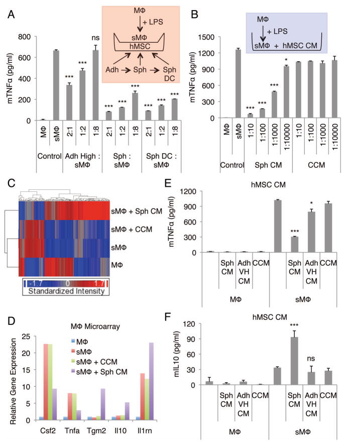
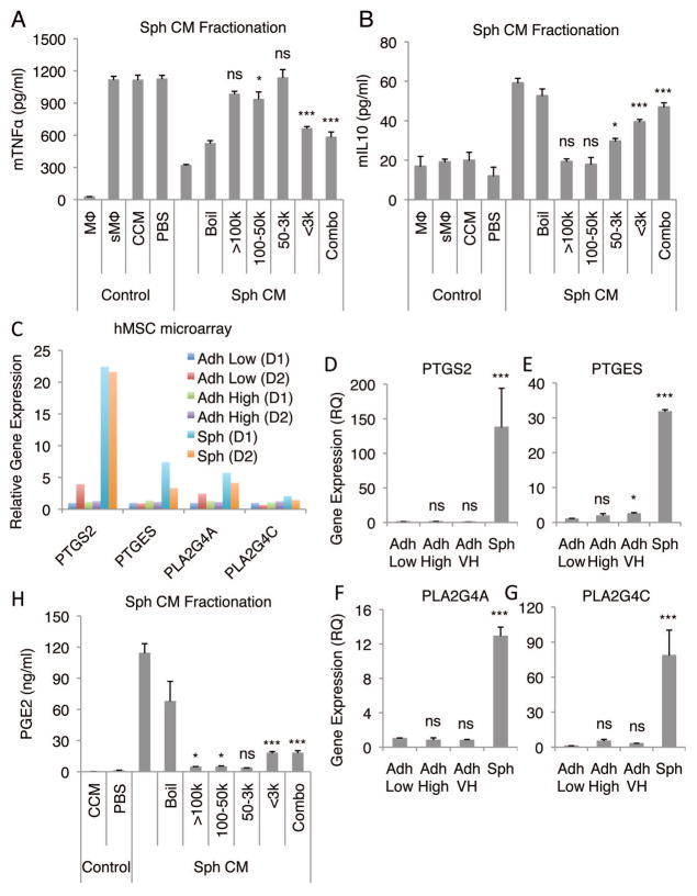
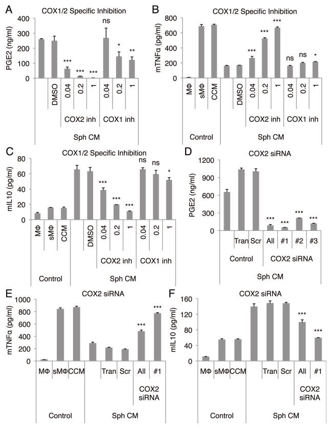
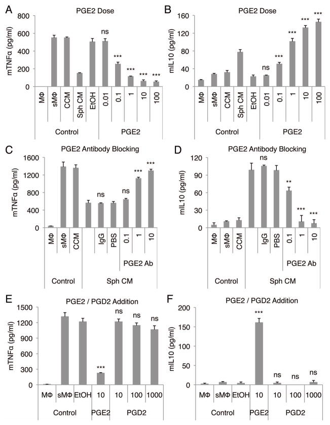
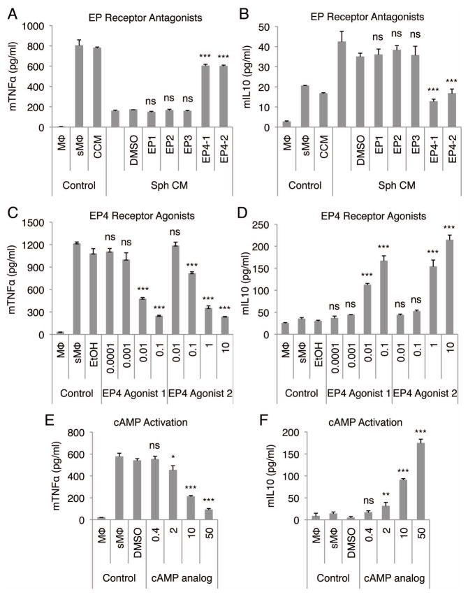
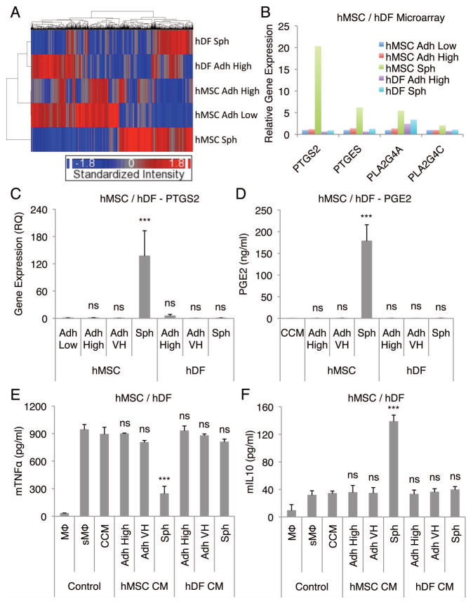
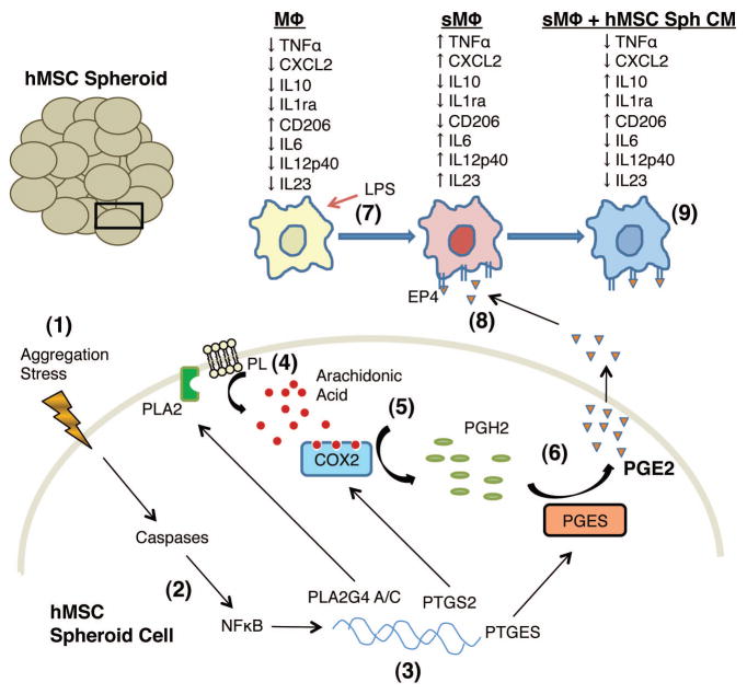
Culturing cells in three dimension (3D) provides an insight into their characteristics in vivo. We previously reported that human mesenchymal stem/stromal cells (hMSCs) cultured as 3D spheroids acquire enhanced anti-inflammatory properties. Here, we explored the effects of hMSC spheroids on macrophages that are critical cells in the regulation of inflammation. Conditioned medium (CM) from hMSC spheroids inhibited lipopolysaccharide-stimulated macrophages from secreting proinflammatory cytokines TNFα, CXCL2, IL6, IL12p40, and IL23. CM also increased the secretion of anti-inflammatory cytokines IL10 and IL1ra by the stimulated macrophages, and augmented expression of CD206, a marker of alternatively activated M2 macrophages. The principal anti-inflammatory activity in CM had a small molecular weight, and microarray data suggested that it was prostaglandin E2 (PGE2). This was confirmed by the observations that PGE2 levels were markedly elevated in hMSC spheroid-CM, and that the anti-inflammatory activity was abolished by an inhibitor of cyclooxygenase-2 (COX-2), a silencing RNA for COX-2, and an antibody to PGE2. The anti-inflammatory effects of the PGE2 on stimulated macrophages were mediated by the EP4 receptor. Spheroids formed by human adult dermal fibroblasts produced low levels of PGE2 and displayed negligible anti-inflammatory effects on stimulated macrophages, suggesting the features as unique to hMSCs. Moreover, production of PGE2 by hMSC spheroids was dependent on the activity of caspases and NFκB activation in the hMSCs. The results indicated that hMSCs in 3D-spheroid cultures are self-activated, in part by intracellular stress responses, to produce PGE2 that can change stimulated macrophages from a primarily proinflammatory M1 phenotype to a more anti-inflammatory M2 phenotype.


Figures

Figure 1
Figure 1. hMSC spheroids and spheroid-conditioned medium promote anti-inflammatory macrophage phenotype
(A) hMSC spheroids and spheroid-derived cells reduce the secretion of mTNFα by LPS-stimulated macrophages. Transwell co-cultures of macrophages and hMSCs at different cell ratios. (B) hMSC spheroid-conditioned medium reduces the secretion of mTNFα by LPS-stimulated macrophages at a dose-dependent manner. (C) Hierarchical clustering of mouse macrophage microarray data. (D) Relative gene expression levels of selected inflammatory related genes from the mouse macrophage microarray data. Un-stimulated mouse macrophages were used as a baseline. (E,F) Comparison of the spheroid and monolayer hMSC conditioned medium effect in production of mTNFα (E) and mIL10 (F) by LPS-stimulated macrophages. Values are mean ± SD (n = 3). ns P ≥ 0.05, *P < 0.05, ***P < 0.001 compared to control (sMΦ) in A, compared to corresponding vehicle controls (CCM) in B, and compared to vehicle control (CCM) in E and F. Abbreviations: Adh High, adherent monolayer hMSCs plated at high density (5,000 cells/cm2) and cultured for 3 d; Adh VH, adherent monolayer hMSCs plated at very high density (200,000 cells/cm2) and cultured for 3 d; CM, conditioned medium; CCM, complete culture medium; hMSC, human mesenchymal stem/stromal cell; LPS, lipopolysaccharide; mTNFα, mouse TNFα; MΦ, macrophage; sMΦ, stimulated macrophage; Sph, spheroid hMSC from 3 d hanging drop cultures (25,000 cells/drop); Sph DC, spheroid-derived cell.
Figure 2
Figure 2. Fractionation of hMSC spheroid-conditioned medium identifies PGE2 as a candidate anti-inflammatory molecule
(A,B) Effects of fractions of hMSC spheroid-conditioned medium on production of mTNFα (A) and mIL10 (B) by LPS-stimulated macrophages. (C) Relative gene expression levels of molecules involved in PGE2 synthesis from microarray data from 3 different preparations of hMSCs from 2 different donors. hMSCs plated at a low density (100–150 cells/cm2) and grown for 7 d were used as a baseline. (D–G) Real-time PCR expression data for PTGS2 (D), PTGES (E), PLA2G4A (F), and PLA2G4C (G) in hMSC monolayers and spheroids. hMSCs plated at a low density and grown for 7 d were used as a baseline. (H) PGE2 content of hMSC spheroid-conditioned medium fractions. Values are mean ± SD (n = 3). ns P ≥ 0.05, *P < 0.05, ***P < 0.001 compared to vehicle control (CCM) in A, B, and H and compared to Adh Low in D, E, F, and G. Abbreviations: Boil, denatured hMSC Sph CM; Combo, equal combination of all the fractions; D1, donor 1; k, kDa; PBS, phosphate buffered saline. Other abbreviations as in Fig. 1.
Figure 3
Figure 3. Anti-inflammatory effect of hMSC spheroid-conditioned medium is dependent on COX-2
(A-C) COX-2 inhibition in hMSC spheroids reduces the production of PGE2 by the hMSCs (A), and the conditioned medium fails to reduce mTNFα (B) and increase mIL10 (C) secretion by the stimulated macrophages. COX-1 and 2 inhibitor concentrations are shown in μM. (D–F) COX-2 knockdown in hMSC spheroids reduces the production of PGE2 by the hMSCs (D), and the conditioned medium fails to reduce mTNFα (E) and increase mIL10 (F) secretion by the stimulated macrophages. Values are mean ± SD (n = 3). ns P ≥ 0.05, *P < 0.05, **P < 0.01, ***P < 0.001 compared to vehicle control (DMSO) in A, B, and C and compared to control (Scr) in D, E, and F. Abbreviations: All, combination of COX-2 siRNA #1, 2, and 3; DMSO, dimethyl sulfoxide; inh, inhibitor; Scr, negative control siRNA; Tran, Transfection reagent control; #1, 2, and 3, three different siRNAs for COX-2. Other abbreviations as in Fig. 1.
Figure 4
Figure 4. Spheroid hMSC produced PGE2 has a strong anti-inflammatory effect on stimulated macrophages
(A,B) PGE2 reduces the secretion of mTNFα (A) and increases the secretion of mIL10 (B) by LPS-stimulated macrophages in a dose-dependent manner. PGE2 doses are shown in ng/ml. (C,D) PGE2 neutralization in hMSC spheroid-conditioned medium reduces the anti-inflammatory effect of the conditioned medium on LPS-stimulated macrophages measured as mTNFα (C) and mIL10 (D) secretion. PGE2 antibody doses are shown in μg/ml. (E,F) PGD2 does not reduce the secretion of mTNFα (E) or increase the secretion of mIL10 (F) by LPS-stimulated macrophages. PGE2 and PGD2 doses are shown in ng/ml. Values are mean ± SD (n = 3). ns P ≥ 0.05, **P < 0.01, ***P < 0.001 compared to vehicle control (EtOH) in A, B, E, and F and compared to control (IgG) in C and D. Abbreviations: Ab, antibody; EtOH, ethanol; PBS, phosphate buffered saline. Other abbreviations as in Fig. 1.
Figure 5
Figure 5. Anti-inflammatory effect of hMSC spheroid-conditioned medium is mediated through EP4 receptor on stimulated macrophages
(A,B) EP4 receptor antagonists inhibit the anti-inflammatory effect of hMSC spheroid-conditioned medium on stimulated macrophages. mTNFα (A) and mIL10 (B) ELISA. EP receptor antagonists were used at 10 μM. (C,D) Two different EP4 receptor agonists decrease the production of mTNFα (C) and increase the production of mIL10 (D) by stimulated macrophages in a dose-dependent manner. Doses are shown in ng/ml. (E,F) cAMP analog decreases the production of mTNFα (E) and increases the production of mIL10 (F) by stimulated macrophages in a dose-dependent manner. Doses are shown in μM. Values are mean ± SD (n = 3). ns P ≥ 0.05, *P < 0.05, **P < 0.01, ***P < 0.001 compared to vehicle control (DMSO) in A, B, E, and F and compared to vehicle control (EtOH) in C and D. Abbreviations: DMSO, dimethyl sulfoxide; EtOH, ethanol. Other abbreviations as in Fig. 1.
Figure 6
Figure 6. hDFs do not produce high levels of PGE2 and do not have an anti-inflammatory effect on stimulated macrophages
(A) Hierarchical clustering of hMSC and hDF microarray data. (B) Relative gene expression levels of molecules involved in PGE2 synthesis from the hMSC and hDF microarray data. hMSCs plated at a low density and grown for 7 d were used as a baseline. (C,D) hDF spheroids do not express high levels of PTGS2 (C) or secrete PGE2 (D). (E,F) Conditioned medium from hDF spheroids does not lower the secretion of mTNFα (E) or increase the secretion of mIL10 (F) by LPS-stimulated macrophages. Values are mean ± SD (n = 3). ns P ≥ 0.05, ***P < 0.001 compared to Adh Low in C and compared to vehicle control (CCM) in D, E, and F. Abbreviation: hDF, human adult dermal fibroblast. Other abbreviations as in Fig. 1.
Figure 7
Figure 7. Schematic of the proposed signaling in spheroid hMSCs promoting anti-inflammatory phenotype in stimulated macrophages
(1) hMSCs aggregate in a hanging drop to form a spheroid. (2) Aggregation and the resulting stress result in activation of caspases and NFκB. (3) The expression of PLA2G4A/C, PTGS2, and PTGES genes is up-regulated. (4) Phospholipases (PLA2) release arachidonic acid from plasma membrane phospholipids (PL). (5) Arachidonic acid is converted into PGH2 by COX-2. (6) PGH2 is converted into PGE2 by PGE synthase (PGES) followed by secretion of PGE2 (7) LPS stimulation of macrophages increases the secretion of pro-inflammatory cytokines TNFα, CXCL2, IL6, IL12p40, and IL23. (8) hMSC spheroid produced PGE2 binds to the EP4 receptor on LPS-stimulated macrophages. (9) PGE2 binding results in decreased secretion of TNFα, CXCL2, IL6, IL12p40, and IL23 and increased secretion of anti-inflammatory cytokines IL10 and IL1ra, and increased surface expression of M2 macrophage marker CD206 by the stimulated macrophages. Abbreviations as in Fig. 1.

Similar articles

Cited by

References

    1. Prockop DJ, Kota DJ, Bazhanov N, et al. Evolving paradigms for repair of tissues by adult stem/progenitor cells (MSCs) J Cell Mol Med. 2010;14:2190–2199. - PMC - PubMed
    1. Stewart DJ, Mei SH. Cell-based therapies for lung vascular diseases: lessons for the future. Proc Am Thorac Soc. 2011;8:535–540. - PubMed
    1. Weiss DJ, Bertoncello I, Borok Z, et al. Stem cells and cell therapies in lung biology and lung diseases. Proc Am Thorac Soc. 2011;8:223–272. - PMC - PubMed
    1. Ranganath SH, Levy O, Inamdar MS, et al. Harnessing the Mesenchymal Stem Cell Secretome for the Treatment of Cardiovascular Disease. Cell Stem Cell. 2012;10:244–258. - PMC - PubMed
    1. Friedenstein AJ, Gorskaja JF, Kulagina NN. Fibroblast precursors in normal and irradiated mouse hematopoietic organs. Exp Hematol. 1976;4:267–274. - PubMed

Publication types

MeSH terms