. 2012 Oct;192(2):397-415.
doi: 10.1534/genetics.112.142570. Epub 2012 Aug 3.

Multiple pathways of duplication formation with and without recombination (RecA) in Salmonella enterica

Affiliations

Multiple pathways of duplication formation with and without recombination (RecA) in Salmonella enterica

Andrew B Reams et al. Genetics. 2012 Oct.

Abstract

Duplications are often attributed to "unequal recombination" between separated, directly repeated sequence elements (>100 bp), events that leave a recombinant element at the duplication junction. However, in the bacterial chromosome, duplications form at high rates (10(-3)-10(-5)/cell/division) even without recombination (RecA). Here we describe 1800 spontaneous lac duplications trapped nonselectively on the low-copy F'(128) plasmid, where lac is flanked by direct repeats of the transposable element IS3 (1258 bp) and by numerous quasipalindromic REP elements (30 bp). Duplications form at a high rate (10(-4)/cell/division) that is reduced only about 11-fold in the absence of RecA. With and without RecA, most duplications arise by recombination between IS3 elements (97%). Formation of these duplications is stimulated by IS3 transposase (Tnp) and plasmid transfer functions (TraI). Three duplication pathways are proposed. First, plasmid dimers form at a high rate stimulated by RecA and are then modified by deletions between IS3 elements (resolution) that leave a monomeric plasmid with an IS3-flanked lac duplication. Second, without RecA, duplications occur by single-strand annealing of DNA ends generated in different sister chromosomes after transposase nicks DNA near participating IS3 elements. The absence of RecA may stimulate annealing by allowing chromosome breaks to persist. Third, a minority of lac duplications (3%) have short (0-36 bp) junction sequences (SJ), some of which are located within REP elements. These duplication types form without RecA, Tnp, or Tra by a pathway in which the palindromic junctions of a tandem inversion duplication (TID) may stimulate deletions that leave the final duplication.


Figures

Figure 1
Figure 1
Duplication by unequal crossing over. This conventional view of duplication formation involves standard recombination events between separated sequence elements embedded in nonhomologous surrounding sequence (top). Recombination between the elements generates a duplication with a copy of the recombining element at the junction between duplicated regions and at the site of a corresponding deletion (middle). Regardless of how a duplication forms, the repeated segment can be lost or further amplified (bottom) by standard RecA-dependent recombination between nonallelic copies at any point of their extensive shared homology (see center).
Figure 2
Figure 2
Duplication by replicative transposition. Replicative transposition can directly create a duplication with a copy of the transposable element at the rearrangement join point. The transposition event generates a novel sequence (circled above) at the juxtaposition of one end of the element and the transpositional target site. This event simultaneously generates a deletion that will appear in a different cell and have a novel sequence at the other end of the junction element. Duplications formed in this way have been observed in this system, but do not explain the involvement of transposase in the duplications described here. Conservative transposition can insert an element copy at a novel site for use by recombinational duplication, but we have not observed such events.
Figure 3
Figure 3
Duplication by single-strand annealing. In this diagram, breaks form at separated sites in different sister chromosomes behind a single replication fork. Breaks may form when a replication fork encounters a nick in its template or by conversion of a simple nick to a double-strand break. In the diagram, these breaks form near sequence repeats. This nicking could be caused by transposase or by fork stalling. The 5′ strands at these breaks are resected, leaving 3′-single-strand extensions that could permit pairing of the repeats. Repair of the paired structure by nucleases and synthesis can lead to a duplication. Simultaneous single strands may coexist at a single collapsed replication fork and allow duplication between nearby repeats as diagramed previously (Lovett et al. 1993) or may form adjacent to transposable elements that are subject to transposase nicking.
Figure 4
Figure 4
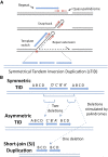
Tandem inversion duplications and their modification. Amplifications of this type were recovered in bacteria and yeast after prolonged growth under selection for additional gene copies (Araya et al. 2010; Kugelberg et al. 2010) and are also found in certain cancer cells, especially following exposure to cancer chemotherapy. Their formation is thought to be initiated by short quasi-palindromic structures. (A) One model for TID formation that relies on initiating repair replication at snap-back structures and switching templates to produce symmetrical triplications whose junctions have extended complementary palindromic sequences. (B) Deletions that modify a sTID to generate asymmetric junctions or simple tandem duplications.
Figure 5
Figure 5
Features of the F′128 plasmid. The thick line represents the portion of the plasmid derived from the F-plasmid. The lac-containing segment between IS3A and IS3C is derived from the E. coli chromosome. Elements IS3A and IS3C are identical and differ from IS3B by seven base substitutions. REP elements are quasipalindromic repeated sequences that fall into three related families (Y, Z1, and Z2). These elements are typically found as clusters with multiple pairs of inversely oriented REP sequences.
Figure 6
Figure 6
Structure of the duplication types. The haploid parent (top) has a lac operon flanked by copies of IS3 and several clusters of palindromic REP elements (see Figure 5). Duplications to form between separated short sequences and leave one copy of the sequence at the join point indicated by parentheses. While these junctions appear to have formed by unequal recombination, all duplication types can form in strains lacking RecA.
Figure 7
Figure 7
Effect of RecA and IS3 on formation of duplications. The frequency of several duplication types was determined for populations grown 33 generations without selection. RecA affects formation of duplications with IS3A/C junctions but not duplications with short junction sequences. Strains lacking RecA still produce an appreciable number of IS3A/C duplications.
Figure 8
Figure 8
Effect of IS3 transposase on lac duplication formation. All strains except the one at the far left have a deletion of the defective IS3B element. For each strain, the two duplication types were determined: IS3 (open columns) and REP plus SJ (red columns). In the third, fourth, and fifth strain, IS3A and IS3C sequences are present but transposase (Tnp) production has been eliminated from one, the other, or both elements by a base substitution in the initiation codon of the tnp gene. In the strain at the far right (shaded column), both IS3A and IS3C were individually replaced by directly oriented copies of the rifR gene, leaving lac flanked by identical sequence repeats with no encoded transposase.
Figure 9
Figure 9
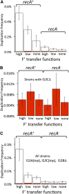
Effects of plasmid transfer functions. (A) The effect of tra expression—and thefore TraI nicking at OriT—on duplication frequency in rec+ and recA strains. (B) The effect of tra expression on the frequency of SJ plus REP duplication types in strains lacking IS3C and therefore incapable of making duplications with an IS3A/C junction. (C) The effect of tra on duplication frequency in strains with flanking IS3 elements but lacking transposase. Throughout the figure, the expression level “none” is a traI mutant with no nicking at OriT, “low” is a traI+ strain with tra operon repressed by pSLT), and “high” is a traI+ strain with derepressed tra operon (no pSLT).
Figure 10
Figure 10
Effects of recB and recF mutations. For each strain, the two general duplication types were determined: IS3 (open columns) and REP plus SJ (red columns).
Figure 11
Figure 11
Pathways for duplication formation and replicative transposition. (A) Three different pathways that form duplications on F′128. Each pathway can be distinguished on the basis of its dependence on RecA, IS3 transposase, and Tra functions. (B) The process of replicative transposition through a co-integrate intermediate. Note the similarity between the resolution event in B and the deletion affecting the plasmid dimer at left side of A.
Figure 12
Figure 12
Assays of deletion formation on F′128. The figure describes the two events assayed to assess formation of deletions between two IS3 elements. The first test, duplication loss, was performed in recA strains, where duplication loss occurs primarily by exchanges between two IS3 elements. The second test, insert loss rate, uses a monomeric F′128 plasmid and determines the rate at which the insert (lac) is lost by exchanges between nonallelic IS3 elements.
Figure 13
Figure 13
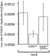
Duplication loss recombination-deficient strains depends on IS3 transposase. The duplication loss rate was measured in recA strains whose F′128 plasmid carries a lac duplication. Three such duplication strains were analyzed: (1) a strain with wild-type IS3 elements, (2) a strain in which all copies of IS3 lack transposase expression, and (3) a strain with impaired IS3 transposase expression and a plasmid that overexpresses IS3 transposase in trans. The frequency of cells without the duplication was measured at various time points during nonselective growth and the duplication loss rates were calculated after correcting for fitness effects using a spreadsheet simulation.
Figure A1
Figure A1
Duplications isolated with and without selection (SJ and REP types). Duplications other that the most frequent IS3C/IS3A type are described in the top half of the figure. The junctions of SJ duplications lie outside of any REP element. These duplications all include lac but their size is not otherwise constrained by the method used to trap them. The REP duplication all arise by exchanges between REP elements lying on either side of lac. The size of these duplications are constrained by the location of REP elements in the F′128 plasmid. The amplification-bearing strains described in the bottom half of the figure were all isolated as Lac+ colonies after prolonged growth under selection on lactose.

Similar articles

Cited by

References

    1. Anderson R. P., Roth J. R., 1978. Tandem chromosomal duplications in Salmonella typhimurium: fusion of histidine genes to novel promoters. J. Mol. Biol. 119: 147–166 - PubMed
    1. Anderson P., Roth J., 1981. Spontaneous tandem genetic duplications in Salmonella typhimurium arise by unequal recombination between rRNA (rrn) cistrons. Proc. Natl. Acad. Sci. USA 78: 3113–3117 - PMC - PubMed
    1. Andersson D. I., Slechta E. S., Roth J. R., 1998. Evidence that gene amplification underlies adaptive mutability of the bacterial lac operon. Science 282: 1133–1135 - PubMed
    1. Andersson D. I., Hughes D., Roth J. R. (), 2011. The Origin of Mutants Under Selection: Interactions of Mutation, Growth, and Selection. ASM Press, Washington, DC. - PubMed
    1. Araya C. L., Payen C., Dunham M. J., Fields S., 2010. Whole-genome sequencing of a laboratory-evolved yeast strain. BMC Genomics 11: 88. - PMC - PubMed

Publication types