

Microtubule-depolymerizing kinesin KLP10A restricts the length of the acentrosomal meiotic spindle in Drosophila females
- PMID: 22865737
- PMCID: PMC3454874
- DOI: 10.1534/genetics.112.143503
Microtubule-depolymerizing kinesin KLP10A restricts the length of the acentrosomal meiotic spindle in Drosophila females
Abstract
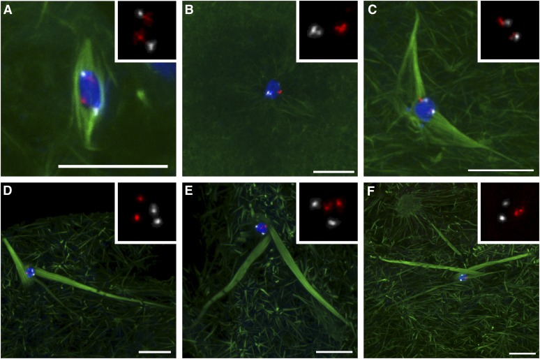
During cell division, a bipolar array of microtubules forms the spindle through which the forces required for chromosome segregation are transmitted. Interestingly, the spindle as a whole is stable enough to support these forces even though it is composed of dynamic microtubules, which are constantly undergoing periods of growth and shrinkage. Indeed, the regulation of microtubule dynamics is essential to the integrity and function of the spindle. We show here that a member of an important class of microtubule-depolymerizing kinesins, KLP10A, is required for the proper organization of the acentrosomal meiotic spindle in Drosophila melanogaster oocytes. In the absence of KLP10A, microtubule length is not controlled, resulting in extraordinarily long and disorganized spindles. In addition, the interactions between chromosomes and spindle microtubules are disturbed and can result in the loss of contact. These results indicate that the regulation of microtubule dynamics through KLP10A plays a critical role in restricting the length and maintaining bipolarity of the acentrosomal meiotic spindle and in promoting the contacts that the chromosomes make with microtubules required for meiosis I segregation.
Figures
Similar articles
-
The chromosomal basis of meiotic acentrosomal spindle assembly and function in oocytes.Chromosoma. 2017 Jun;126(3):351-364. doi: 10.1007/s00412-016-0618-1. Epub 2016 Nov 11. Chromosoma. 2017. PMID: 27837282 Free PMC article. Review.
-
Multiple motors cooperate to establish and maintain acentrosomal spindle bipolarity in C. elegans oocyte meiosis.Elife. 2022 Feb 11;11:e72872. doi: 10.7554/eLife.72872. Elife. 2022. PMID: 35147496 Free PMC article.
-
Cooperation Between Kinesin Motors Promotes Spindle Symmetry and Chromosome Organization in Oocytes.Genetics. 2017 Feb;205(2):517-527. doi: 10.1534/genetics.116.194647. Epub 2016 Dec 7. Genetics. 2017. PMID: 27932541 Free PMC article.
-
Meiosis-specific stable binding of augmin to acentrosomal spindle poles promotes biased microtubule assembly in oocytes.PLoS Genet. 2013 Jun;9(6):e1003562. doi: 10.1371/journal.pgen.1003562. Epub 2013 Jun 13. PLoS Genet. 2013. PMID: 23785300 Free PMC article.
-
Molecular mechanisms of kinesin-14 motors in spindle assembly and chromosome segregation.J Cell Sci. 2017 Jul 1;130(13):2097-2110. doi: 10.1242/jcs.200261. J Cell Sci. 2017. PMID: 28668932 Review.
Cited by
-
Suppression of Patronin deficiency by altered Hippo signaling in Drosophila organ development.Cell Death Differ. 2021 Jan;28(1):233-250. doi: 10.1038/s41418-020-0597-x. Epub 2020 Jul 31. Cell Death Differ. 2021. PMID: 32737445 Free PMC article.
-
The developmental biology of kinesins.Dev Biol. 2021 Jan 1;469:26-36. doi: 10.1016/j.ydbio.2020.09.009. Epub 2020 Sep 19. Dev Biol. 2021. PMID: 32961118 Free PMC article. Review.
-
Female Meiosis: Synapsis, Recombination, and Segregation in Drosophila melanogaster.Genetics. 2018 Mar;208(3):875-908. doi: 10.1534/genetics.117.300081. Genetics. 2018. PMID: 29487146 Free PMC article. Review.
-
The chromosomal basis of meiotic acentrosomal spindle assembly and function in oocytes.Chromosoma. 2017 Jun;126(3):351-364. doi: 10.1007/s00412-016-0618-1. Epub 2016 Nov 11. Chromosoma. 2017. PMID: 27837282 Free PMC article. Review.
-
14-3-3 regulation of Ncd reveals a new mechanism for targeting proteins to the spindle in oocytes.J Cell Biol. 2017 Oct 2;216(10):3029-3039. doi: 10.1083/jcb.201704120. Epub 2017 Aug 31. J Cell Biol. 2017. PMID: 28860275 Free PMC article.
References
-
- Albertson D. G., Thomson J. N., 1993. Segregation of holocentric chromosomes at meiosis in the nematode, Caenorhabditis elegans. Chromosome Res. 1: 15–26 - PubMed
-
- Brand A. H., Perrimon N., 1993. Targeted gene expression as a means of altering cell fates and generating dominant phenotypes. Development 118: 401–415 - PubMed
-
- Casso D., Ramirez-Weber F., Kornberg T. B., 2000. GFP-tagged balancer chromosomes for Drosophila melanogaster. Mech. Dev. 91: 451–454 - PubMed
Publication types
MeSH terms
Substances
Grants and funding
LinkOut - more resources
Full Text Sources
Molecular Biology Databases
