

Carcinomatous meningitis: Yet another cause for rapidly progressive dementia and triphasic waves in electroencephalograph!
- PMID: 22865984
- PMCID: PMC3410003
- DOI: 10.4103/0976-3147.98253
Carcinomatous meningitis: Yet another cause for rapidly progressive dementia and triphasic waves in electroencephalograph!
Abstract
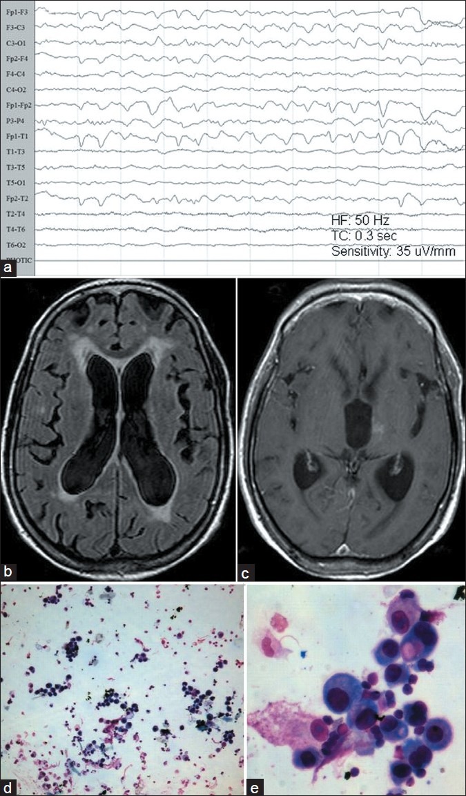
We report a 65-year-old woman who manifested with progressive cognitive impairment, abnormal behavior, slurred speech, inability to carry out activities with right upper limb, gait disturbances, emotional liability, and double incontinence that evolved progressively over the last 8 months. A clinical syndrome of "rapidly progressive dementia" was considered. The MRI of brain was unremarkable except for small para third ventricular enhancing lesion was detected in the left thalamic region. There was bi/tri-phasic sharp waves in the routine scalp EEG occurring at periodically 1.5-2.0 Hz, mimicking Creutzfeldt-Jakob disease (CJD). She was later diagnosed to have carcinomatous meningitis based on cerebrospinal fluid (CSF) cytology. This case is being discussed for rarity and interesting EEG observations in patients with carcinomatous meningitis and to highlight the importance of CSF cytology in an appropriate clinical setting. One needs to be careful in concluding CJD as possible diagnosis in such scenario.
Keywords: Carcinomatous meningitis; dementia; triphasic waves.
Conflict of interest statement
Figures
Similar articles
-
Triphasic waves in electroencephalogram as a possible early marker of carcinomatous meningitis: a case report.Medicine (Baltimore). 2020 Aug 14;99(33):e21735. doi: 10.1097/MD.0000000000021735. Medicine (Baltimore). 2020. PMID: 32872059 Free PMC article.
-
Sporadic onset Creutzfeldt-Jacob disease: interesting MRI observations.Neurol India. 2006 Dec;54(4):418-20. doi: 10.4103/0028-3886.28119. Neurol India. 2006. PMID: 17114856
-
[A 64-year-old woman with progressive gait disturbance and dementia for one year].No To Shinkei. 1998 Sep;50(9):861-70. No To Shinkei. 1998. PMID: 9789311 Japanese.
-
Sporadic Creutzfeldt-Jakob disease with tau pathology mimicking new-onset refractory non-convulsive status epilepticus: Case report and review of the literature.Eur J Neurol. 2021 Apr;28(4):1385-1391. doi: 10.1111/ene.14624. Epub 2020 Nov 27. Eur J Neurol. 2021. PMID: 33135248 Review.
-
Creutzfeldt-Jakob and Vascular Brain Diseases: Their Overlap and Relationships.Front Neurol. 2021 Feb 25;12:613991. doi: 10.3389/fneur.2021.613991. eCollection 2021. Front Neurol. 2021. PMID: 33732205 Free PMC article. Review.
Cited by
-
Commentary.J Neurosci Rural Pract. 2012 May;3(2):210-1. J Neurosci Rural Pract. 2012. PMID: 22865985 Free PMC article. No abstract available.
-
Triphasic waves in electroencephalogram as a possible early marker of carcinomatous meningitis: a case report.Medicine (Baltimore). 2020 Aug 14;99(33):e21735. doi: 10.1097/MD.0000000000021735. Medicine (Baltimore). 2020. PMID: 32872059 Free PMC article.
-
Neurology Case Report: Rapidly Progressive Dementia and Extrapyramidal Symptoms as the First Presentation of Leptomeningeal Carcinomatosis.Cureus. 2022 Mar 7;14(3):e22923. doi: 10.7759/cureus.22923. eCollection 2022 Mar. Cureus. 2022. PMID: 35281578 Free PMC article.
-
Describing Symptom Burden and Functional Status at the Diagnosis of Leptomeningeal Metastasis.Oncol Nurs Forum. 2018 May 1;45(3):372-379. doi: 10.1188/18.ONF.372-379. Oncol Nurs Forum. 2018. PMID: 29683126 Free PMC article.
References
-
- Aguglia U, Gambardella A, Oliveri RL, Lavano A, Quattrone A. Non-metabolic causes of triphasic waves: A reappraisal. Clin Electroencephalogr. 1990;21:120–5. - PubMed
-
- Vas CJ, Mallya MV, Desai M, Mahadevan A, Nadkarni NS, Shankar SK. Carcinomatous meningitis mimicking Creutzfeldt-Jakob disease. Neurol India. 2004;52:383–6. - PubMed
-
- Liss L. Correlation between clinical diagnosis of Jakob-Creutzfeldt disease autopsy findings. Abstract 606 D09. XIV, World Congress of Neurology. Neurol India. 1989;37:360.
-
- Zeidler M, Sellar RJ, Collie DA, Knight R, Stewart G, Macleod MA, et al. The pulvinar sign on magnetic resonance imaging in variant Creutzfeldt-Jakob disease. Lancet. 2000;355:1412–8. - PubMed
Publication types
LinkOut - more resources
Full Text Sources