. 2011 Dec;2(12):1117-33.
doi: 10.1177/1947601911423654.

Hypoxia-induced angiogenesis: good and evil

Affiliations

Hypoxia-induced angiogenesis: good and evil

Bryan L Krock et al. Genes Cancer. 2011 Dec.

Abstract

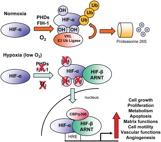
The vascular network delivers oxygen (O(2)) and nutrients to all cells within the body. It is therefore not surprising that O(2) availability serves as a primary regulator of this complex organ. Most transcriptional responses to low O(2) are mediated by hypoxia-inducible factors (HIFs), highly conserved transcription factors that control the expression of numerous angiogenic, metabolic, and cell cycle genes. Accordingly, the HIF pathway is currently viewed as a master regulator of angiogenesis. HIF modulation could provide therapeutic benefit for a wide array of pathologies, including cancer, ischemic heart disease, peripheral artery disease, wound healing, and neovascular eye diseases. Hypoxia promotes vessel growth by upregulating multiple pro-angiogenic pathways that mediate key aspects of endothelial, stromal, and vascular support cell biology. Interestingly, recent studies show that hypoxia influences additional aspects of angiogenesis, including vessel patterning, maturation, and function. Through extensive research, the integral role of hypoxia and HIF signaling in human disease is becoming increasingly clear. Consequently, a thorough understanding of how hypoxia regulates angiogenesis through an ever-expanding number of pathways in multiple cell types will be essential for the identification of new therapeutic targets and modalities.

Keywords: HIFs; angiogenesis; anti-angiogenic therapies; cancer; hypoxia; vascular diseases.


Conflict of interest statement

Declaration of Conflicting Interests: The author(s) declared no potential conflicts of interest with respect to the research, authorship, and/or publication of this article.

Figures

Figure 1.
Figure 1.
Schematic representation of HIF-α and HIF-β structures and DNA binding. (A) Hypoxia-inducible factors (HIFs) are heterodimers composed of 2 subunits: one α (HIF-1α, HIF-2α, HIF-3α) and one β (HIF-β/ARNT). Each subunit contains different functional domains implicated in nuclear localization (NLS), DNA binding (bHLH/PAS), dimerization (bHLH/PAS), protein stability (ODDD/N-TAD), cofactor interactions (N-TAD/C-TAD), and transcriptional activity (N-TAD/C-TAD). NLS = nuclear localization signal; bHLH = basic helix loop helix domain; PAS = Per-ARNT-Sim motif; ODDD = oxygen-dependent degradation domain; N-TAD = N-terminal transactivation domain; C-TAD = C-terminal transactivation domain. For simplicity, only relevant amino acid residues for HIF-1α and HIF-β are represented. However, distinct residues have been identified for HIF-2α. Hydroxylated residues (red), phosphorylated residues (green), sumoylated residues (blue), nitrosylated residues (gray), and acetylated residues (pink). (B) Active HIF complex binds specific hypoxia-responsive elements (HRE, consensus sequence: G/ACGTG) in the promoters of target genes with the basic residues near the N-terminal terminus of each subunit and induces transcription with co-activators bound through the transactivation domains (TAD) of each protein.
Figure 2.
Figure 2.
Regulation of hypoxia-inducible factor (HIF) activity. Under normoxic conditions, HIF-α subunits are polyubiquitinated at 2 proline residues within the oxygen-dependent degradation domain (ODDD) by a family of enzymes known as prolyl hydroxylases (PHDs). This promotes recognition by the VHL E3 ubiquitin ligase complex and subsequent degradation of HIF-α via the 26S proteasome. In addition, hydroxylation of a C-terminal asparagine residue of HIF-α by factor-inhibiting HIF-1 (FIH-1) prevents binding of cofactors required for HIF activity. Hypoxia inhibits the activity of the PHD and FIH-1 enzymes, allowing HIF-α proteins to escape recognition by VHL, be stabilized, and translocate to the nucleus. There, they dimerize with HIF-1β/ARNT and bind hypoxia response elements (HREs) within the promoters of target genes. Together with the co-activator proteins p300 and CBP, the HIF complex activates the transcription of a panel of genes required for the response to hypoxia. OH = hydroxylation; Ub = ubiquitin.
Figure 3.
Figure 3.
Regulation of angiogenesis and angiogenic steps by oxygen availability through the hypoxia-inducible factor (HIF)–induced angiogenic factors. It is now well established that, in response to hypoxia, HIFs regulate angiogenic genes (see Table 2) to control angiogenic steps and functions in embryonic vascular development and pathologic settings such as cancer or vascular diseases. CACs = circulating angiogenic cells; EPCs = endothelial progenitor cells.
Figure 4.
Figure 4.
Determining the complex effect of local hypoxia on pathological angiogenesis is likely to have important therapeutic consequences. Since downregulation of the hypoxia-inducible factors (HIFs) inhibits angiogenesis and decreases tumor growth, regulation of the expression and activity of HIFs is evolving as a potential target for modulating tumor angiogenesis and is becoming an attractive approach for the treatment of solid tumors and macular degeneration, maladies typically associated with unregulated or excessive angiogenic activity. Inhibitors of HIF synthesis, stability, dimerization, and DNA binding are currently in clinical trials and show promising results. In contrast, forced expression or activation of HIFs, using viral vectors or prolyl hydroxylase (PHD) inhibitors, could evolve as a potential solution for the improvement of therapeutic angiogenesis and revascularization after critical ischemia in adults affected by ischemic diseases. The ability to induce and regulate angiogenesis and vascular remodeling in a directed manner would represent a major advance in the treatment of ischemic vascular diseases, including myocardial infarction, atherosclerosis, and peripheral artery disease. HDAC = histone deacetylase; CAC = circulating angiogenic cell.

References

    1. Semenza GL. Life with oxygen. Science. 2007; 318:62-4 - PubMed
    1. Carmeliet P. Angiogenesis in life, disease and medicine. Nature. 2005;438:932-6 - PubMed
    1. Bertout JA, Patel SA, Simon MC. The impact of O2 availability on human cancer. Nat Rev Cancer. 2008;8:967-75 - PMC - PubMed
    1. Carreau A, Hafny-Rahbi BE, Matejuk A, Grillon C, Kieda C. Why is the partial oxygen pressure of human tissues a crucial parameter? Small molecules and hypoxia. J Cell Mol Med. 2011;15:1239-53 - PMC - PubMed
    1. Parmar K, Mauch P, Vergilio JA, Sackstein R, Down JD. Distribution of hematopoietic stem cells in the bone marrow according to regional hypoxia. Proc Natl Acad Sci U S A. 2007;104:5431-6 - PMC - PubMed

LinkOut - more resources