. 2012 Aug 6:9:31.
doi: 10.1186/1743-8977-9-31.

Glutathione-S-transferase M1 regulation of diesel exhaust particle-induced pro-inflammatory mediator expression in normal human bronchial epithelial cells

Affiliations

Glutathione-S-transferase M1 regulation of diesel exhaust particle-induced pro-inflammatory mediator expression in normal human bronchial epithelial cells

Weidong Wu et al. Part Fibre Toxicol. .

Abstract

Background: Diesel exhaust particles (DEP) contribute substantially to ambient particulate matter (PM) air pollution in urban areas. Inhalation of PM has been associated with increased incidence of lung disease in susceptible populations. We have demonstrated that the glutathione S-transferase M1 (GSTM1) null genotype could aggravate DEP-induced airway inflammation in human subjects. Given the critical role airway epithelial cells play in the pathogenesis of airway inflammation, we established the GSTM1 deficiency condition in primary bronchial epithelial cells from human volunteers with GSTM1 sufficient genotype (GSTM1+) using GSTM1 shRNA to determine whether GSTM1 deficiency could exaggerate DEP-induced expression of interleukin-8 (IL-8) and IL-1β proteins. Furthermore, the mechanisms underlying GSTM1 regulation of DEP-induced IL-8 and IL-1β expression were also investigated.

Methods: IL-8 and IL-1β protein levels were measured using enzyme-linked immunosorbent assay. GSTM1 deficiency in primary human bronchial epithelial cells was achieved using lentiviral GSTM1 shRNA particles and verified using real-time polymerase chain reaction and immunoblotting. Intracellular reactive oxygen species (ROS) production was evaluated using flow cytometry. Phosphorylation of protein kinases was detected using immunoblotting.

Results: Exposure of primary human bronchial epithelial cells (GSTM1+) to 25-100 μg/ml DEP for 24 h significantly increased IL-8 and IL-1β protein expression. Knockdown of GSTM1 in these cells further elevated DEP-induced IL-8 and IL-1β expression, implying that GSTM1 deficiency aggravated DEP-induced pro-inflammatory response. DEP stimulation induced the phosphorylation of extracellular signal-regulated kinase (ERK) and Akt, the downstream kinase of phosphoinositide 3-kinase (PI3K), in GSTM1+ bronchial epithelial cells. Pharmacological inhibition of ERK kinase and PI3K activity blocked DEP-induced IL-8 and IL-1β expression. DEP-induced ERK and Akt phosphorylation could be increased by GSTM1 knockdown. In addition, pretreatment of HBEC with the antioxidant N-acetyl cysteine significantly inhibited DEP-induced ERK and Akt phosphorylation, and subsequent IL-8 and IL-1β expression.

Conclusion: GSTM1 regulates DEP-induced IL-8 and IL-1β expression in primary human bronchial epithelial cells by modulation of ROS, ERK and Akt signaling.


Figures
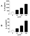
Figure 1
Figure 1
DEP exposure increases IL-8 and IL-1β protein expression in HBEC. Confluent HBEC were exposed to control (PBS) or 25–100 μg/ml DEP for 24 h, respectively. IL-8 and IL-1β levels in the supernatants of culture media were measured using ELISA.
Figure 2
Figure 2
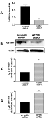
GSTM1knockdown enhanced DEP-induced IL-8 and IL-1β expression in HBEC. A and B, HBEC were infected with lentiviral scrambled or GSTM1 shRNA particles (10 moi) for 24 h, respectively. The cells were lysed for GSTM1 mRNA measurement using RT-PCR (*P < 0.01)or GSTM1 protein determination using immunoblotting. Infected HBEC with lentiviral scrambled or GSTM1 shRNA particles were treated with PBS control or 50 μg/ml DEP for 24 h. Levels of IL-8 (C) and IL-1β (D) proteins in the supernatant of culture media were determined using ELISA and expressed as fold over control (DEP vs. PBS control in scrambled or GSTM1 shRNA group, respectively). *P < 0.05, compared to DEP treatment in the scrambled shRNA group.
Figure 3
Figure 3
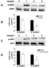
The ERK and the PI3K/Akt pathways are involved in DEP-induced IL-8 and IL-1β expression.A, Confluent HBEC were exposed to 50 μg/ml DEP for 1-4 h. Supernatants of cell lysates were subjected to SDS–PAGE and immunoblotting using phospho- and pan antibodies against ERK. B, HBEC were pretreated with vehicle (DMSO), or 20 μM ERK kinase inhibitor U0126, for 30 min prior to 50 μg/ml DEP stimulation for 24 h, respectively. Levels of IL-8 and IL-1β proteins were determined using ELISA and expressed as fold over control. *P < 0.05, compared to DEP treatment in the vehicle (DMSO) group. C, Confluent HBEC were exposed to 50 μg/ml DEP for 1-4 h. Phosphorylated Akt was determined using immunoblotting and phospho-specific antibody against Akt. D, HBEC were pretreated with vehicle (DMSO), or 1 μM PI3K inhibitor Wortmannin, for 30 min prior to 50 μg/ml DEP stimulation for 24 h, respectively. Levels of IL-8 and IL-1β proteins were measured using ELISA and expressed as fold over control (DEP vs. PBS control in vehicle DMSO or inhibitor group, respectively). * P < 0.05, compared to DEP treatment in the vehicle (DMSO) group.
Figure 4
Figure 4
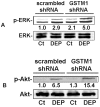
GSTM1knockdown enhances DEP-induced ERK and Akt activation. HBEC were infected with lentiviral scrambled or GSTM1 shRNA particles (10 moi) for 24 h, respectively. Confluent cells were treated with PBS control (Ct) or 50 μg/ml DEP for 1 h. The supernatants of the cell lysates were subjected to immunoblotting using phospho- or pan antibodies against ERK (A) or Akt (B), respectively. Shown are representative bands derived from 4 separate experiments. Nos. are mean relative optical densities of phorspho-proteins compared to control.
Figure 5
Figure 5
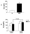
GSTM1knockdown enhances DEP-induced ROS production.A, HBEC were treated with 50 μg/ml DEP for 1 h. Intracellular ROS levels were measured using the fluorescent probe carboxy-H2DCFDA and flow cytometry. B, HBEC were infected with 10 moi of lentiviral non-target (scrambled) or GSTM1 shRNA particles for 24 h, respectively. Confluent cells were treated with 50 μg/ml DEP for 4 h and ROS levels measured as described previously. *, compared to PBS control (Ct), P < 0.05.
Figure 6
Figure 6
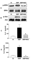
NAC reduced DEP-induced ERK and Akt phosphorylation, and IL-8 and IL-1β expression.A and B, HBEC were pretreated with 10 mM NAC for 2 h before further stimulation with 50 μg/ml DEP for 1 h. Phosphorylated ERK and Akt was measured using imunoblotting. C and D, HBEC were pretreated with 10 mM NAC for 2 h before further stimulation with 50 μg/ml DEP for 24 h. Levels of IL-8 and IL-1β proteins were measured using ELISA. *, compared to DEP, P < 0.05.

References

    1. Gowdy K, Krantz QT, Daniels M, Linak WP, Jaspers I, Gilmour MI. Modulation of pulmonary inflammatory responses and antimicrobial defenses in mice exposed to diesel exhaust. Toxicol Appl Pharmacol. 2008;229:310–319. doi: 10.1016/j.taap.2008.01.040. - DOI - PubMed
    1. Saxon A, Diaz-Sanchez D. Air pollution and allergy: you are what you breathe. Nat Immunol. 2005;6:223–226. doi: 10.1038/ni0305-223. - DOI - PubMed
    1. McCreanor J, Cullinan P, Nieuwenhuijsen MJ, Stewart-Evans J, Malliarou E, Jarup L, Harrington R, Svartengren M, Han IK, Ohman-Strickland P, Chung KF, Zhang J. Respiratory effects of exposure to diesel traffic in persons with asthma. N Engl J Med. 2007;357:2348–2358. doi: 10.1056/NEJMoa071535. - DOI - PubMed
    1. Ris C. U.S. EPA health assessment for diesel engine exhaust: a review. Inhal Toxicol. 2007;19(Suppl 1):229–239. - PubMed
    1. Raphael GD, Metcalfe DD. Mediators of airway inflammation. Eur J Respir Dis Suppl. 1986;147:44–56. - PubMed

Publication types

MeSH terms