

Elevated expression of the chemokine-scavenging receptor D6 is associated with impaired lesion development in psoriasis
- PMID: 22867710
- PMCID: PMC3532592
- DOI: 10.1016/j.ajpath.2012.06.042
Elevated expression of the chemokine-scavenging receptor D6 is associated with impaired lesion development in psoriasis
Abstract
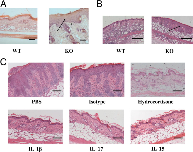
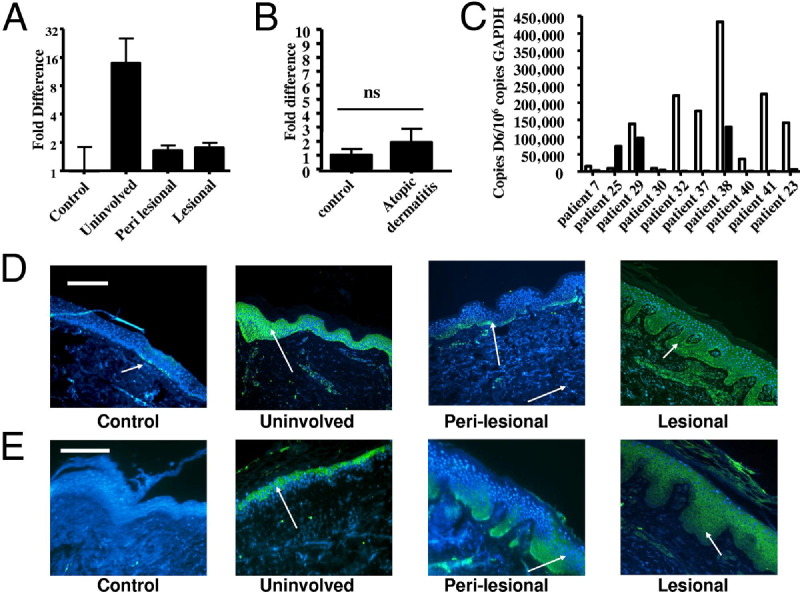
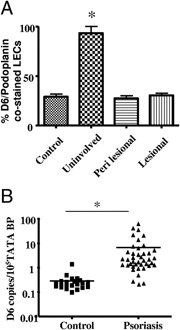
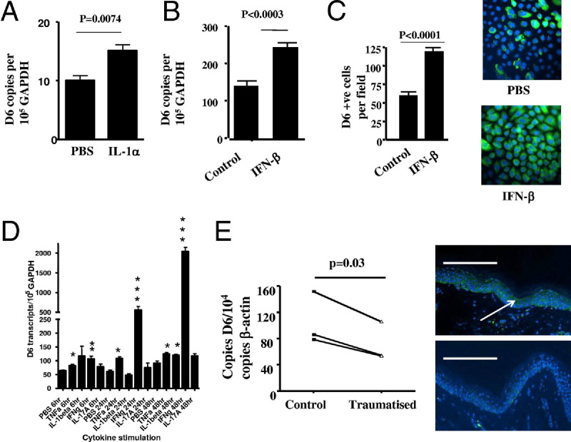
D6 is a scavenging-receptor for inflammatory CC chemokines that are essential for resolution of inflammatory responses in mice. Here, we demonstrate that D6 plays a central role in controlling cutaneous inflammation, and that D6 deficiency is associated with development of a psoriasis-like pathology in response to varied inflammatory stimuli in mice. Examination of D6 expression in human psoriatic skin revealed markedly elevated expression in both the epidermis and lymphatic endothelium in "uninvolved" psoriatic skin (ie, skin that was more than 8 cm distant from psoriatic plaques). Notably, this increased D6 expression is associated with elevated inflammatory chemokine expression, but an absence of plaque development, in uninvolved skin. Along with our previous observations of the ability of epidermally expressed transgenic D6 to impair cutaneous inflammatory responses, our data support a role for elevated D6 levels in suppressing inflammatory chemokine action and lesion development in uninvolved psoriatic skin. D6 expression consistently dropped in perilesional and lesional skin, coincident with development of psoriatic plaques. D6 expression in uninvolved skin also was reduced after trauma, indicative of a role for trauma-mediated reduction in D6 expression in triggering lesion development. Importantly, D6 is also elevated in peripheral blood leukocytes in psoriatic patients, indicating that upregulation may be a general protective response to inflammation. Together our data demonstrate a novel role for D6 as a regulator of the transition from uninvolved to lesional skin in psoriasis.
Copyright © 2012 American Society for Investigative Pathology. Published by Elsevier Inc. All rights reserved.
Figures
Similar articles
-
Microarray analyses demonstrate the involvement of type I interferons in psoriasiform pathology development in D6-deficient mice.J Biol Chem. 2013 Dec 20;288(51):36473-83. doi: 10.1074/jbc.M113.491563. Epub 2013 Nov 5. J Biol Chem. 2013. PMID: 24194523 Free PMC article.
-
Cutaneous lesions in psoriatic arthritis are enriched in chemokine transcriptomic pathways.Arthritis Res Ther. 2023 May 2;25(1):73. doi: 10.1186/s13075-023-03034-6. Arthritis Res Ther. 2023. PMID: 37131254 Free PMC article.
-
An analysis of the function and expression of D6 on lymphatic endothelial cells.Blood. 2013 May 2;121(18):3768-77. doi: 10.1182/blood-2012-04-425314. Epub 2013 Mar 11. Blood. 2013. PMID: 23479571
-
Regulation of the immune and inflammatory responses by the 'atypical' chemokine receptor D6.J Pathol. 2013 Jan;229(2):168-75. doi: 10.1002/path.4123. J Pathol. 2013. PMID: 23125030 Review.
-
D6 and the atypical chemokine receptor family: novel regulators of immune and inflammatory processes.Eur J Immunol. 2009 Feb;39(2):342-51. doi: 10.1002/eji.200838858. Eur J Immunol. 2009. PMID: 19130487 Review.
Cited by
-
Cell-autonomous regulation of neutrophil migration by the D6 chemokine decoy receptor.J Immunol. 2013 Jun 15;190(12):6450-6456. doi: 10.4049/jimmunol.1201429. Epub 2013 May 13. J Immunol. 2013. PMID: 23670187 Free PMC article.
-
Elevated ACKR2 expression is a common feature of inflammatory arthropathies.Rheumatology (Oxford). 2017 Sep 1;56(9):1607-1617. doi: 10.1093/rheumatology/kex176. Rheumatology (Oxford). 2017. PMID: 28486662 Free PMC article.
-
CXCL10 Is an Agonist of the CC Family Chemokine Scavenger Receptor ACKR2/D6.Cancers (Basel). 2021 Mar 2;13(5):1054. doi: 10.3390/cancers13051054. Cancers (Basel). 2021. PMID: 33801414 Free PMC article.
-
Spread of Psoriasiform Inflammation to Remote Tissues Is Restricted by the Atypical Chemokine Receptor ACKR2.J Invest Dermatol. 2017 Jan;137(1):85-94. doi: 10.1016/j.jid.2016.07.039. Epub 2016 Aug 26. J Invest Dermatol. 2017. PMID: 27568525 Free PMC article.
-
MicroRNA-146 and cell trauma down-regulate expression of the psoriasis-associated atypical chemokine receptor ACKR2.J Biol Chem. 2018 Feb 23;293(8):3003-3012. doi: 10.1074/jbc.M117.809780. Epub 2017 Dec 26. J Biol Chem. 2018. PMID: 29279330 Free PMC article.
References
-
- Nickoloff B.J., Qin J.Z., Nestle F.O. Immunopathogenesis of psoriasis. Clin Rev Allergy Immunol. 2007;33:45–56. - PubMed
-
- van de Kerkhof P.C. The evolution of the psoriatic lesion. Br J Dermatol. 2007;157:4–15. - PubMed
-
- Gudjonsson J.E., Ding J., Li X., Nair R.P., Tejasvi T., Qin Z.S., Ghosh D., Aphale A., Gumucio D.L., Voorhees J.J., Abecasis G.R., Elder J.T. Global gene expression analysis reveals evidence for decreased lipid biosynthesis and increased innate immunity in uninvolved psoriatic skin. J Invest Dermatol. 2009;129:2795–2804. - PMC - PubMed
-
- Rot A., von Andrian U.H. Chemokines in innate and adaptive host defense: basic chemokinese grammar for immune cells. Annu Rev Immunol. 2004;22:891–928. - PubMed
-
- Homey B., Meller S. Chemokines and other mediators as therapeutic targets in psoriasis vulgaris. Clin Dermatol. 2008;26:539–545. - PubMed
Publication types
MeSH terms
Substances
Grants and funding
LinkOut - more resources
Full Text Sources
Medical
Molecular Biology Databases
