

Rheumatoid and pyrophosphate arthritis synovial fibroblasts induce osteoclastogenesis independently of RANKL, TNF and IL-6
- PMID: 22867712
- PMCID: PMC3593104
- DOI: 10.1016/j.jaut.2012.06.001
Rheumatoid and pyrophosphate arthritis synovial fibroblasts induce osteoclastogenesis independently of RANKL, TNF and IL-6
Abstract
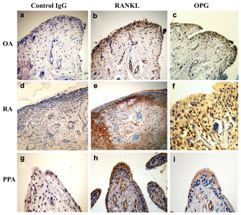
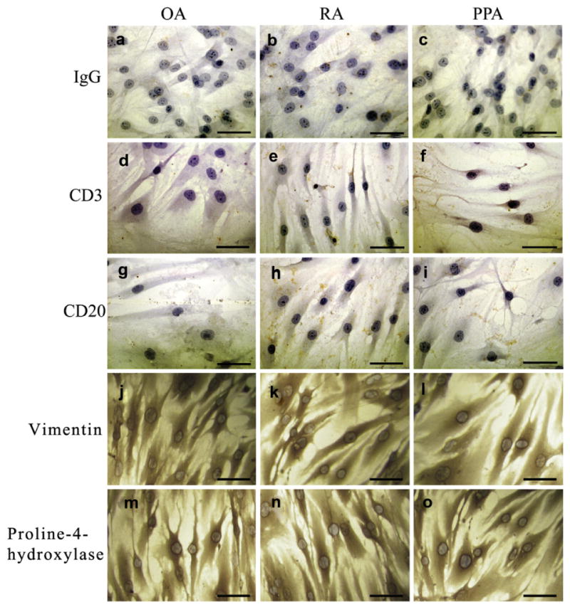
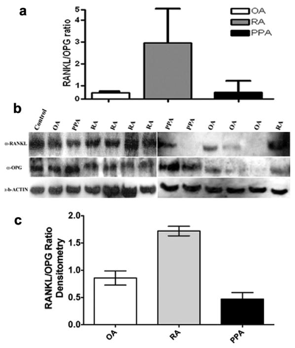
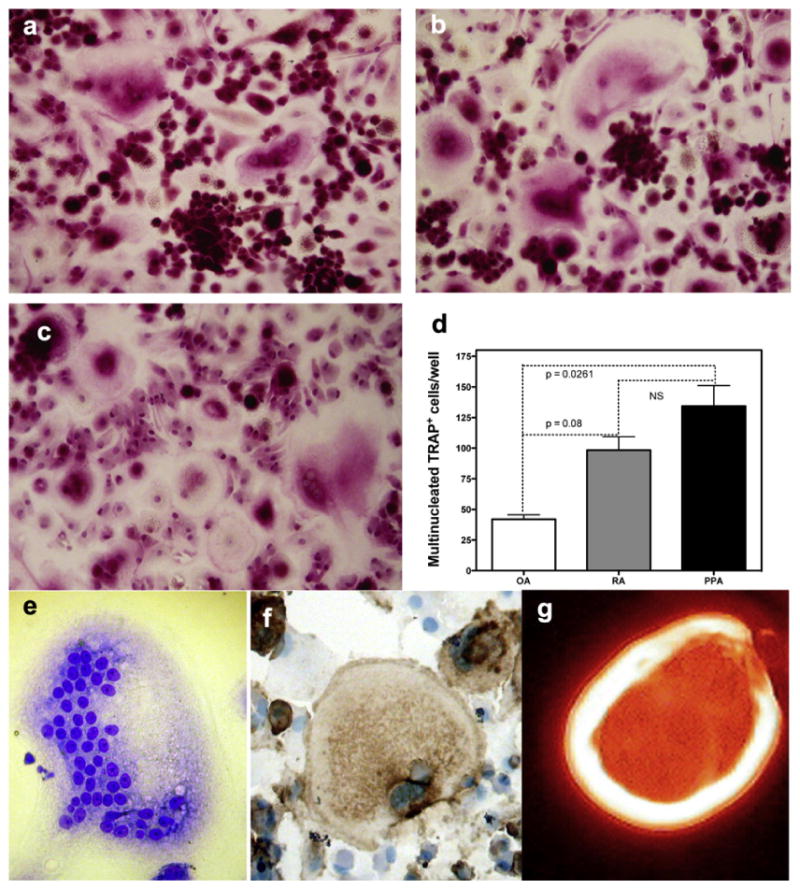
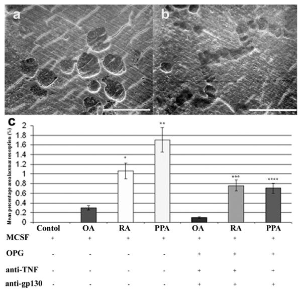
Bone destruction is a common feature of inflammatory arthritis and is mediated by osteoclasts, the only specialized cells to carry out bone resorption. Aberrant expression of receptor activator of nuclear factor kappa β ligand (RANKL), an inducer of osteoclast differentiation has been linked with bone pathology and the synovial fibroblast in rheumatoid arthritis (RA). In this manuscript, we challenge the current concept that an increase in RANKL expression governs osteoclastogenesis and bone destruction in autoimmune arthritis. We isolated human fibroblasts from RA, pyrophosphate arthropathy (PPA) and osteoarthritis (OA) patients and analyzed their RANKL/OPG expression profile and the capacity of their secreted factors to induce osteoclastogenesis. We determined a 10-fold increase of RANKL mRNA and protein in fibroblasts isolated from RA relative to PPA and OA patients. Peripheral blood mononuclear cells (PBMC) from healthy volunteers were cultured in the presence of RA, PPA and OA synovial fibroblast conditioned medium. Osteoclast differentiation was assessed by expression of tartrate-resistant acid phosphatase (TRAP), vitronectin receptor (VNR), F-actin ring formation and bone resorption assays. The formation of TRAP(+), VNR(+) multinucleated cells, capable of F-actin ring formation and lacunar resorption in synovial fibroblast conditioned medium cultures occured in the presence of osteoprotegerin (OPG) a RANKL antagonist. Osteoclasts did not form in these cultures in the absence of macrophage colony stimulating factor (M-CSF). Our data suggest that the conditioned medium of pure synovial fibroblast cultures contain inflammatory mediators that can induce osteoclast formation in human PBMC independently of RANKL. Moreover inhibition of the TNF or IL-6 pathway was not sufficient to abolish osteoclastogenic signals derived from arthritic synovial fibroblasts. Collectively, our data clearly show that alternate osteoclastogenic pathways exist in inflammatory arthritis and place the synovial fibroblast as a key regulatory cell in bone and joint destruction, which is a hallmark of autoimmune arthritis.
Copyright © 2012 Elsevier Ltd. All rights reserved.
Figures
Comment in
-
Bone: An alternative pathway for bone destruction in inflammatory arthritis?Nat Rev Rheumatol. 2012 Oct;8(10):563. doi: 10.1038/nrrheum.2012.141. Epub 2012 Aug 21. Nat Rev Rheumatol. 2012. PMID: 22907291 No abstract available.
References
-
- Muller-Ladner U, Gay RE, Gay S. Activation of synoviocytes. Curr Opin Rheumatol. 2000;12:186–94. - PubMed
Publication types
MeSH terms
Substances
Grants and funding
LinkOut - more resources
Full Text Sources
Medical
Research Materials
