

Deletion of heart-type cytochrome c oxidase subunit 7a1 impairs skeletal muscle angiogenesis and oxidative phosphorylation
- PMID: 22869013
- PMCID: PMC3497574
- DOI: 10.1113/jphysiol.2012.239707
Deletion of heart-type cytochrome c oxidase subunit 7a1 impairs skeletal muscle angiogenesis and oxidative phosphorylation
Abstract
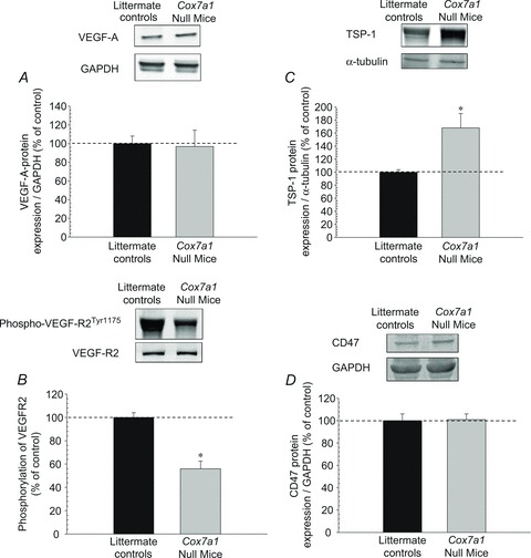
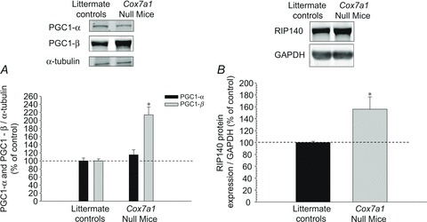
Oxidative metabolism is needed for sustained skeletal muscle function. A key component of such metabolism is cytochrome c oxidase, the 13-subunit terminal complex of the mitochondrial electron transport chain. We used mice null for one of the two isoforms of Cox subunit 7a, heart/skeletal muscle-specific Cox7a1, to examine the cellular and functional responses of muscle adaptation in response to mitochondrial dysfunction. Specifically we determined if deletion of Cox7a1 would (1) limit exercise capacity, and (2) alter genes responsible for skeletal muscle capillarity and mitochondrial biogenesis. Sixteen male mice (Cox7a1 null mice, n = 8, and littermate controls, n = 8) performed incremental and run-to-exhaustion treadmill tests. The hindlimb muscles for both groups were analysed. The results indicated that capillary indices were reduced (by 30.7–44.9%) in the Cox7a1 null mice relative to controls. In addition, resting ATP levels and Cox specific activity were significantly reduced (>60%) in both glycolytic and oxidative muscle fibre types despite an increase in a major regulator of mitochondrial biogenesis, PGC-1β. These changes in the skeletal muscle resulted in exercise intolerance for the Cox7a1 null mice. Thus, our data indicate that deletion of the Cox7a1 isoform results in reduced muscle bioenergetics and hindlimb capillarity, helping to explain the observed impairment of muscle structure and function.
Figures


Similar articles
-
Adult skeletal muscle deletion of Mitofusin 1 and 2 impedes exercise performance and training capacity.J Appl Physiol (1985). 2019 Feb 1;126(2):341-353. doi: 10.1152/japplphysiol.00719.2018. Epub 2018 Sep 27. J Appl Physiol (1985). 2019. PMID: 30260752 Free PMC article.
-
Mice deleted for heart-type cytochrome c oxidase subunit 7a1 develop dilated cardiomyopathy.Mitochondrion. 2012 Mar;12(2):294-304. doi: 10.1016/j.mito.2011.11.002. Epub 2011 Nov 20. Mitochondrion. 2012. PMID: 22119795 Free PMC article.
-
Global deletion of thrombospondin-1 increases cardiac and skeletal muscle capillarity and exercise capacity in mice.Exp Physiol. 2009 Jun;94(6):749-60. doi: 10.1113/expphysiol.2008.045989. Epub 2009 Mar 18. Exp Physiol. 2009. PMID: 19297388
-
Aerobic Exercise Preconception and During Pregnancy Enhances Oxidative Capacity in the Hindlimb Muscles of Mice Offspring.J Strength Cond Res. 2018 May;32(5):1391-1403. doi: 10.1519/JSC.0000000000002416. J Strength Cond Res. 2018. PMID: 29309390 Free PMC article.
-
Tissue-specific adaptations to cytochrome c oxidase deficiency shape physiological outcomes.Biochim Biophys Acta Mol Basis Dis. 2025 Mar;1871(3):167567. doi: 10.1016/j.bbadis.2024.167567. Epub 2024 Nov 28. Biochim Biophys Acta Mol Basis Dis. 2025. PMID: 39613003 Review.
Cited by
-
Mitochondrial Diseases Part I: mouse models of OXPHOS deficiencies caused by defects in respiratory complex subunits or assembly factors.Mitochondrion. 2015 Mar;21:76-91. doi: 10.1016/j.mito.2015.01.009. Epub 2015 Feb 4. Mitochondrion. 2015. PMID: 25660179 Free PMC article. Review.
-
Considerations for aerobic exercise paradigms with rodent models.Lab Anim (NY). 2016 May 20;45(6):213-5. doi: 10.1038/laban.1023. Lab Anim (NY). 2016. PMID: 27203261 No abstract available.
-
Plasma proteomics show altered inflammatory and mitochondrial proteins in patients with neurologic symptoms of post-acute sequelae of SARS-CoV-2 infection.Brain Behav Immun. 2023 Nov;114:462-474. doi: 10.1016/j.bbi.2023.08.022. Epub 2023 Sep 11. Brain Behav Immun. 2023. PMID: 37704012 Free PMC article.
-
COX7A1 suppresses the viability of human non-small cell lung cancer cells via regulating autophagy.Cancer Med. 2019 Dec;8(18):7762-7773. doi: 10.1002/cam4.2659. Epub 2019 Oct 30. Cancer Med. 2019. PMID: 31663688 Free PMC article.
-
Tissue- and Condition-Specific Isoforms of Mammalian Cytochrome c Oxidase Subunits: From Function to Human Disease.Oxid Med Cell Longev. 2017;2017:1534056. doi: 10.1155/2017/1534056. Epub 2017 May 16. Oxid Med Cell Longev. 2017. PMID: 28593021 Free PMC article. Review.
References
-
- Acin-Perez R, Bayona-Bafaluy MP, Bueno M, Machicado C, Fernandez-Silva P, Perez-Martos A, et al. An intragenic suppressor in the cytochrome c oxidase I gene of mouse mitochondrial DNA. Hum Mol Genet. 2003;12:329–339. - PubMed
-
- Arany Z, Foo SY, Ma Y, Ruas JL, Bommi-Reddy A, Girnun G, et al. HIF-independent regulation of VEGF and angiogenesis by the transcriptional coactivator PGC-1α. Nature. 2008;451:1008–1012. - PubMed
-
- Arany Z, Lebrasseur N, Morris C, Smith E, Yang W, Ma Y, Chin S, Spiegelman BM. The transcriptional coactivator PGC-1β drives the formation of oxidative type IIX fibers in skeletal muscle. Cell Metab. 2007;5:35–46. - PubMed
Publication types
MeSH terms
Substances
Grants and funding
LinkOut - more resources
Full Text Sources
Other Literature Sources
Molecular Biology Databases