. 2012 Jul;3(7):709-22.
doi: 10.18632/oncotarget.588.

Frequent ATRX, CIC, FUBP1 and IDH1 mutations refine the classification of malignant gliomas

Affiliations

Frequent ATRX, CIC, FUBP1 and IDH1 mutations refine the classification of malignant gliomas

Yuchen Jiao et al. Oncotarget. 2012 Jul.

Abstract

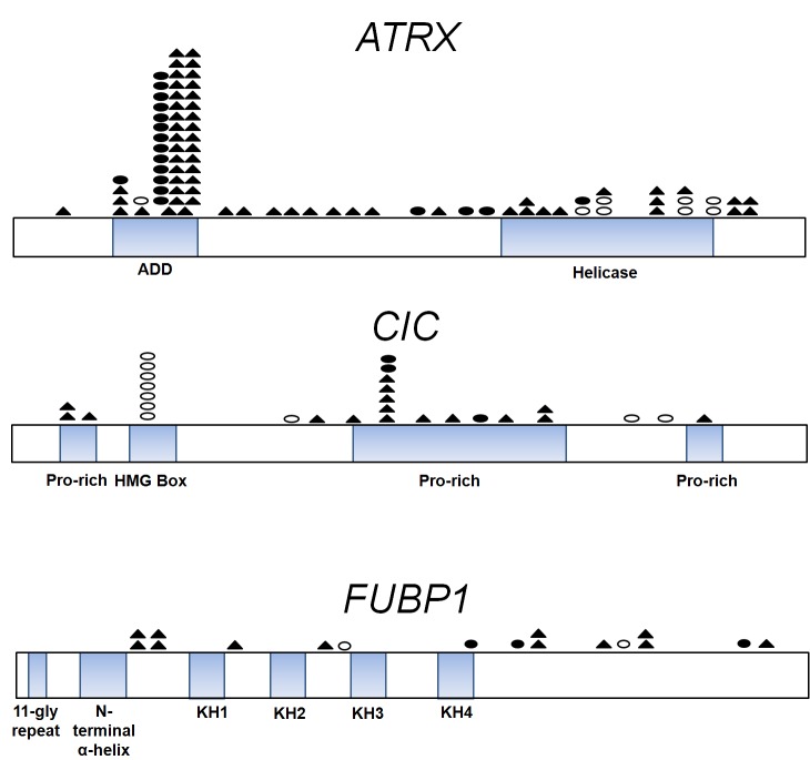
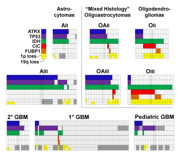
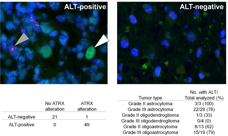
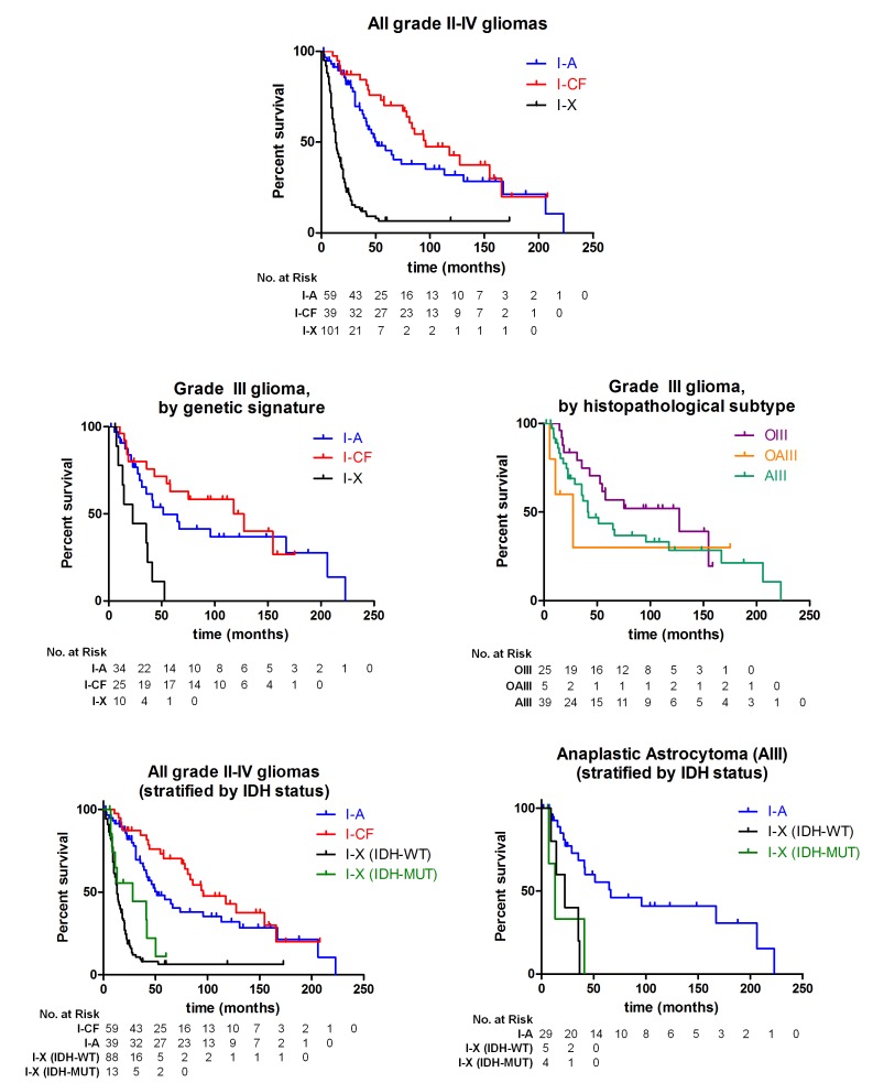
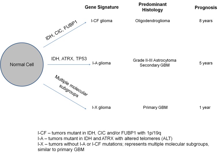
Mutations in the critical chromatin modifier ATRX and mutations in CIC and FUBP1, which are potent regulators of cell growth, have been discovered in specific subtypes of gliomas, the most common type of primary malignant brain tumors. However, the frequency of these mutations in many subtypes of gliomas, and their association with clinical features of the patients, is poorly understood. Here we analyzed these loci in 363 brain tumors. ATRX is frequently mutated in grade II-III astrocytomas (71%), oligoastrocytomas (68%), and secondary glioblastomas (57%), and ATRX mutations are associated with IDH1 mutations and with an alternative lengthening of telomeres phenotype. CIC and FUBP1 mutations occurred frequently in oligodendrogliomas (46% and 24%, respectively) but rarely in astrocytomas or oligoastrocytomas ( more than 10%). This analysis allowed us to define two highly recurrent genetic signatures in gliomas: IDH1/ATRX (I-A) and IDH1/CIC/FUBP1 (I-CF). Patients with I-CF gliomas had a significantly longer median overall survival (96 months) than patients with I-A gliomas (51 months) and patients with gliomas that did not harbor either signature (13 months). The genetic signatures distinguished clinically distinct groups of oligoastrocytoma patients, which usually present a diagnostic challenge, and were associated with differences in clinical outcome even among individual tumor types. In addition to providing new clues about the genetic alterations underlying gliomas, the results have immediate clinical implications, providing a tripartite genetic signature that can serve as a useful adjunct to conventional glioma classification that may aid in prognosis, treatment selection, and therapeutic trial design.


Figures

Figure 1
Figure 1. Distribution of ATRX, CIC, and FUBP1 mutations in gliomas
Schematics of the coding sequences of ATRX, CIC, and FUBP1 are shown. Mutations were predominantly truncating mutations such as frameshifts (shaded triangles) and nonsense mutations (shaded circles). Empty circles correspond to missense mutations. ATRX contains a DNA-binding ATRX-DNMT3-DNMT3L (ADD) domain and an ATPase Helicase Domain. ATRX alterations occurred in 20% of pediatric GBMs (n=25) and all of those mutations clustered in the Helicase domain of ATRX as described previously [14]. ATRX mutations in adult gliomas instead distributed evenly. CIC contains four highly conserved domains, the HMG (high mobility group) box responsible for DNA binding and proline-rich (pro-rich) domains. Among CIC mutations (n=29), a predilection for missense mutations occurred in the HMG box domain (24%). Mutations in FUBP1 (n=16) occurred 3' of the N-terminal α-helix domain, which controls binding of FUBP1 to the FBP-interacting repressor (FIR). Gene representations are to scale.
Figure 2
Figure 2. Distribution of ATRX, TP53, IDH, CIC, and FUBP1 mutations, and of chromosomes 1p and 19q loss, in grade II-IV gliomas
Data are from 15 grade II astrocytomas (AII), 44 grade III astrocytomas (AIII), 21 grade II oligodendrogliomas (OII), 29 grade III oligodendrogliomas (OIII), 18 grade II oligoastrocytomas (OAII), 22 grade III oligoastrocytomas (OAIII), 14 secondary GBMs (2° GBM), 94 primary GBMs (1° GBM), and 25 pediatric GBMs. chromosome 1p status is indicated in the top of the lower row, and chromosome19q status is indicated in the bottom of this cell, with yellow coloring indicating loss of the indicated chromosomal arm. Gray cells denote analyses which were not informative or for which additional genetic material was not available for analysis.
Figure 3
Figure 3. ALT associates with the ATRX alterations and the I-A signature
(A) representative grade III astrocytoma section that was positive for ALT is shown on the left. A nucleus demonstrating large, ultra-bright telomeres (pink) is indicated with the shaded arrow. A p53 positive nucleus (green), which reflects p53 dysfunction, is indicated with the empty arrow. A representative image of a grade III astrocytoma section that was negative for both ALT and for p53 nuclear staining is shown on the right. The number of tumors with ALT and/or ATRX alterations is shown, along with the percentage of each tumor type with ALT. All tumors with ATRX alterations were I-A, that is, they also had an IDH mutation (n=50); the remaining tumors were IDH wild-type and did not have an ATRX alteration (n=21).
Figure 4
Figure 4. Survival of glioma patients with I-A, I-CF, and I-X genetic signatures
Kaplan-Meier estimates of survival are shown for adult glioma patients, with the number of patients at risk at each time point for each group shown below the respective plot. Among all adult patients with grade II-IV gliomas (n=199), those with I-A tumors and with I-CF tumors survived significantly longer (median 51 and 96 months) than patients with I-X tumors (13 months; P <0.001 for both comparisons). Among grade III glioma patients with tumors of all histologies (n=69), there was a significant difference in survival between groups of patients with I-A or I-CF tumors and patients with I-X tumors (I-A and I-X, P = 0.002; I-CF and I-X, P <0.001). There was not a significant difference in survival when the same grade III glioma patients were stratified by the histopathological diagnosis of their tumors (P = 0.37). When all 199 grade II-IV gliomas were additionally stratified by IDH status, patients with I-A tumors and patients with I-CF tumors still survived significantly longer (median 51 and 96 months) than patients with IDH-mutated I-X tumors, and also longer than patients with IDH-WT I-X tumors (median 28 and 13 months, respectively; P <0.001 for all four comparisons). When grade III astrocytoma patients (n=39) were stratified by genetic signature and IDH status of their tumors, patients with I-A tumors survived longer (66 months) than grade III astrocytoma patients with either IDH-mutated I-X tumors (22 months, P = 0.005) and patients with IDH-WT I-X tumors (13 months, P = 0.002).
Figure 5
Figure 5. Model for molecular classification of gliomas
Alterations in IDH and ATRX comprise the I-A signature, and alterations in IDH as well as one or more of CIC, FUBP1, or combined 1p/19q loss comprise the I-CF signature. I-X gliomas do not have either signature and likely represent multiple molecular disease subgroups. The I-A and I-CF signatures arise early in the pathogenesis of a glioma from a normal precursor cell. I-CF gliomas are typically grade II-III oligodendrogliomas and patients with these tumors survive for about 8 years. I-A gliomas are typically grade II-III astrocytomas or grade IV secondary GBMs and patients with these tumors survive for about 5 years. I-X gliomas are typically grade IV primary GBMs and patients with these tumors survive for about 1 year.

References

    1. Louis DN, Ohgaki H, Wiestler OD, Cavenee WK, Burger PC, Jouvet A, Scheithauer BW, Kleihues P. The 2007 WHO classification of tumours of the central nervous system. Acta neuropathologica. 2007;114(2):97–109. - PMC - PubMed
    1. Wen PY, Kesari S. Malignant glioma in adults (vol 359, pg 492, 2008) New Engl J Med. 2008;359(8):877–877. - PubMed
    1. Ohgaki H, Kleihues P. Genetic alterations and signaling pathways in the evolution of gliomas. Cancer Sci. 2009;100(12):2235–2241. - PMC - PubMed
    1. Cancer Genome Atlas Research N. Comprehensive genomic characterization defines human glioblastoma genes and core pathways. Nature. 2008;455(7216):1061–1068. - PMC - PubMed
    1. Parsons DW, Jones S, Zhang XS, Lin JCH, Leary RJ, Angenendt P, Mankoo P, Carter H, Siu IM, Gallia GL, Olivi A, McLendon R, Rasheed BA, Keir S, Nikolskaya T, Nikolsky Y, et al. An integrated genomic analysis of human glioblastoma Multiforme. Science. 2008;321(5897):1807–1812. - PMC - PubMed

Publication types

MeSH terms