. 2012 Sep 28;287(40):33179-90.
doi: 10.1074/jbc.M111.337063. Epub 2012 Aug 6.

Osterix regulates calcification and degradation of chondrogenic matrices through matrix metalloproteinase 13 (MMP13) expression in association with transcription factor Runx2 during endochondral ossification

Affiliations

Osterix regulates calcification and degradation of chondrogenic matrices through matrix metalloproteinase 13 (MMP13) expression in association with transcription factor Runx2 during endochondral ossification

Riko Nishimura et al. J Biol Chem. .

Abstract

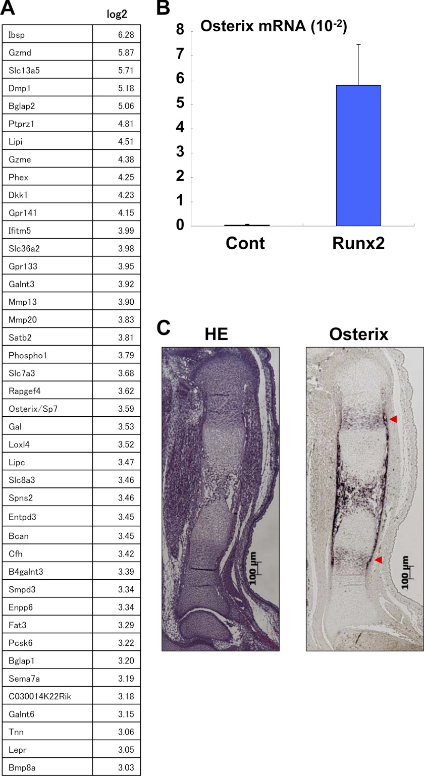
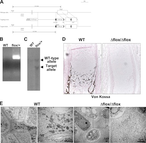
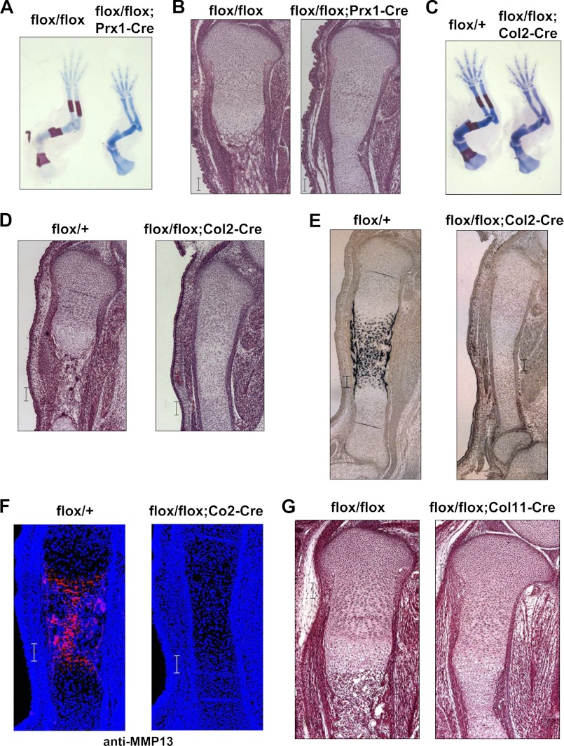
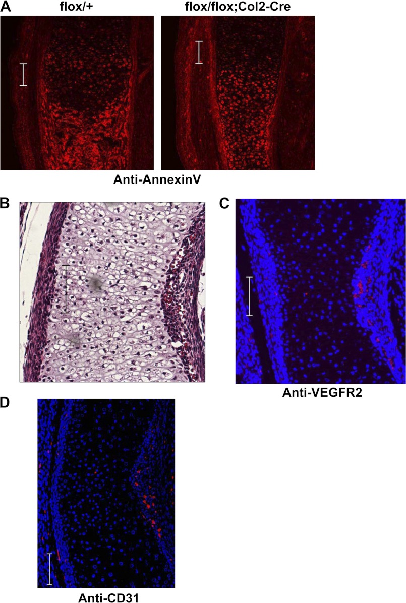
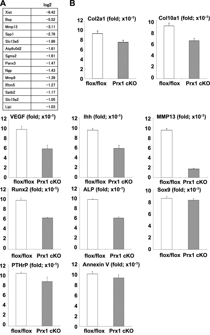
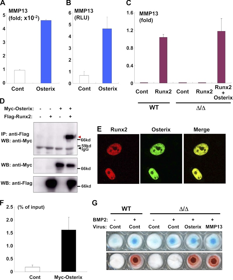
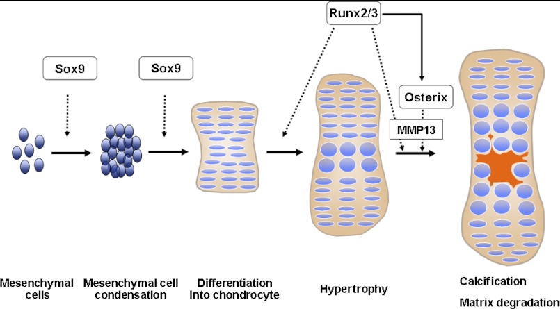
Endochondral ossification is temporally and spatially regulated by several critical transcription factors, including Sox9, Runx2, and Runx3. Although the molecular mechanisms that control the late stages of endochondral ossification (e.g. calcification) are physiologically and pathologically important, these precise regulatory mechanisms remain unclear. Here, we demonstrate that Osterix is an essential transcription factor for endochondral ossification that functions downstream of Runx2. The global and conditional Osterix-deficient mice studied here exhibited a defect of cartilage-matrix ossification and matrix vesicle formation. Importantly, Osterix deficiencies caused the arrest of endochondral ossification at the hypertrophic stage. Microarray analysis revealed that matrix metallopeptidase 13 (MMP13) is an important target of Osterix. We also showed that there exists a physical interaction between Osterix and Runx2 and that these proteins function cooperatively to induce MMP13 during chondrocyte differentiation. Most interestingly, the introduction of MMP13 stimulated the calcification of matrices in Osterix-deficient mouse limb bud cells. Our results demonstrated that Osterix was essential to endochondral ossification and revealed that the physical and functional interaction between Osterix and Runx2 were necessary for the induction of MMP13 during endochondral ossification.


Figures

FIGURE 1.
FIGURE 1.
Up-regulation by Runx2 and expression in cartilage of Osterix. A, mouse limb bud cells were infected with control (Cont) or the Runx2 adenovirus, and then the RNA isolated from the cells was determined by microarray analysis. Up-regulated genes by Runx2 overexpression are listed. B, up-regulation of Osterix expression in mouse limb bud cells by Runx2 was determined using real-time PCR. Values represent means ± S.D. C, Osterix expression was determined by HE staining and in situ hybridization. Hypertrophic-positive areas are indicated by red arrows. Scale bar, 100 μm.
FIGURE 2.
FIGURE 2.
Impaired ossification and matrix vesicle formation in Osterix knock-out mice. A, schematic representation of the Osterix-floxed allele. B, PCR analysis of Osterix-floxed mice. C, heterozygous Osterix-floxed mice were determined by Southern blotting analysis. D, Von Kossa staining of tibia at E16.5 days. WT, wild-type littermate: Δflox/Δflox, Osterix global knock-out mice. Scale bar, 100 μm. E, electron microscopy images of hypertrophic chondrocyte zone of WT and Osterix global knock-out mice (Δflox/Δflox). Right panels show higher magnification. Scale bar, 2 μm. Pr Neo, promoter neomycin resistant gene; Pr DT-A, promoter diphtheria toxin fragment A.
FIGURE 3.
FIGURE 3.
Impairment of ossification in Osterix conditional mice. A and B, flox/+;Prx1-Cre transgenic mice were mated with flox/flox mice. Skeletal preparation of forelimb stained with alcian blue and alizarin red at E15.5 days. Osterix conditional knock-out (flox/flox;Prx1-Cre) and control littermate (flox/flox) (A) are shown. HE staining of tibia of Osterix-conditional knock-out and control littermate at E16.5 days are shown (B). C–E, flox/+;Col2a1(Col2)-Cre transgenic mice were mated with flox/flox mice. Skeletal preparation of forelimb stained with alcian blue and alizarin red at E16.5 days. Osterix-conditional knock-out (flox/flox;Col2-Cre) and control littermate (flox/+) are shown (C). HE staining of tibia of Osterix-conditional knock-out and control littermate at E16.5 days (D). Von Kossa staining of tibia of Osterix-conditional knock-out and control littermate at E16.5 (E). F, MMP13 immunostaining of tibia of Osterix-conditional knock-out and control littermate at E16.5 days. G, flox/+;Col11a2(Col11)-Cre transgenic mice were mated with flox/flox mice. HE staining of tibia of Osterix-conditional knock-out (flox/flox;Col11-Cre) and control littermate (flox/flox) at E16.5 days. Scale bar, 100 μm.
FIGURE 4.
FIGURE 4.
Apoptosis and angiogenesis in Osterix knock-out mice. A, flox/+ and flox/flox;Col2-Cre littermate mice were immunostained with anti-annexin V antibody of tibia of Osterix-conditional knock-out and control littermate at E16.5 days. B, paraffin section of Osterix-conditional knock-out mice (flox/flox;Co2-Cre) at E16.5 days was determined by HE staining. Angiogenesis around the diaphysis of the bone collar was observed in Osterix-conditional knock-out mice (flox/flox;Co2-Cre). C, histological section of Osterix-conditional knock-out mice (flox/flox;Co2-Cre) at E16.5 days was stained with anti-VEGFR2 antibody (red) and DAPI. D, histological section of Osterix-conditional knock-out mice (flox/flox;Co2-Cre) at E16.5 days was stained with anti-CD31 antibody (red) and DAPI. Scale bar, 100 μm.
FIGURE 5.
FIGURE 5.
Down-regulation of MMP13 expression in Osterix-conditional knock-out mice. A, microarray analysis of limb bud tissues of Osterix-conditional knock-out mice (flox/flox;Prx1-Cre) and control littermates from E16.5 days. Down-regulated genes in Osterix-conditional knock-out are listed. B, real-time PCR analyses of limb bud tissues of Osterix-conditional knock-out mice (Prx1 cKO) and control littermates (flox/flox). Values represent means ± S.D.
FIGURE 6.
FIGURE 6.
Osterix and Runx2 required for up-regulation of MMP13. A, up-regulation of MMP13 by Osterix was determined by real-time PCR. Limb bud cells isolated from Osterix knock-out mice were infected with control or Osterix adenovirus, and RNA was isolated from the cells. Values represent means ± S.D. B, ATDC5 cells were transfected with the MMP13 gene promoter luciferase construct and then infected with control (Cont) or Osterix adenovirus. Luciferase activity in the cell lysates was measured. Values represent means ± S.D. C, limb bud cells of wild-type or Osterix knock-out mice (Δ/Δ) were infected with control, Runx2, and/or Osterix adenovirus, and RNA was subjected to real-time PCR analysis. Values represent means ± S.D. D, co-immunoprecipitation experiment using nuclear extracts containing 293 cells transfected with FLAG-Runx2 and/or Myc-Osterix. Co-immunoprecipitated Myc-Osterix with FLAG-Runx2 is shown by the red arrow. IP, immunoprecipitation; WB, Western blotting. E, 293 cells were transfected with DsRed-tagged-Runx2 and Venus-tagged-Osterix and then monitored under confocal microscopy. F, ATDC5 cells infected control (Cont) or Myc-Osterix adenovirus were subjected to a ChIP assay. Immunoprecipitated chromatin samples with anti-Myc antibody and input samples were determined by real-time PCR analysis using a specific Taqman probe against the ∼500 bp upstream region of the MMP13 gene promoter. G, limb bud cells of wild-type or Osterix knock-out mice (Δ/Δ) were subjected to micromass culture. The cells were infected with control, Osterix, or both MMP13 and Cre adenovirus and were subsequently cultured in the presence or absence of BMP2 for 7 days; cells were then stained with alcian blue (top panel) or alizarin red (lower panel).
FIGURE 7.
FIGURE 7.
Schematic diagram of endochondral ossification. Endochondral ossification is sequentially regulated by Sox9, Runx2, and Osterix. Osterix functions as both a downstream and transcriptional partner of Runx2/3 during calcification and matrix degradation in cartilage, and cooperation between Osterix and Runx2/3 are required for MMP13 expression.

References

    1. Kronenberg H. M. (2003) Developmental regulation of the growth plate. Nature 423, 332–336 - PubMed
    1. de Crombrugghe B., Lefebvre V., Behringer R. R., Bi W., Murakami S., Huang W. (2000) Transcriptional mechanisms of chondrocyte differentiation. Matrix Biol. 19, 389–394 - PubMed
    1. Komori T. (2011) Signaling networks in RUNX2-dependent bone development. J. Cell. Biochem. 112, 750–755 - PubMed
    1. Nishimura R., Hata K., Ikeda F., Ichida F., Shimoyama A., Matsubara T., Wada M., Amano K., Yoneda T. (2008) Signal transduction and transcriptional regulation during mesenchymal cell differentiation. J. Bone Miner. Metab. 26, 203–212 - PubMed
    1. Ornitz D. M., Marie P. J. (2002) FGF signaling pathways in endochondral and intramembranous bone development and human genetic disease. Genes Dev. 16, 1446–1465 - PubMed

Publication types