. 2012 Sep 28;287(40):33268-81.
doi: 10.1074/jbc.M112.368142. Epub 2012 Aug 6.

Estrogen facilitates spinal cord synaptic transmission via membrane-bound estrogen receptors: implications for pain hypersensitivity

Affiliations

Estrogen facilitates spinal cord synaptic transmission via membrane-bound estrogen receptors: implications for pain hypersensitivity

Yan Zhang et al. J Biol Chem. .

Abstract

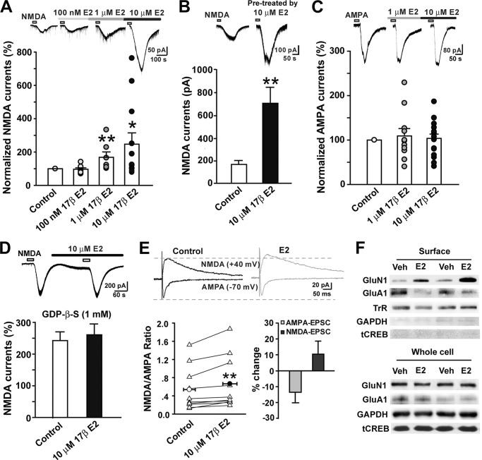
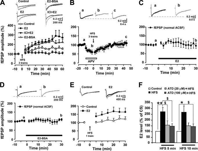
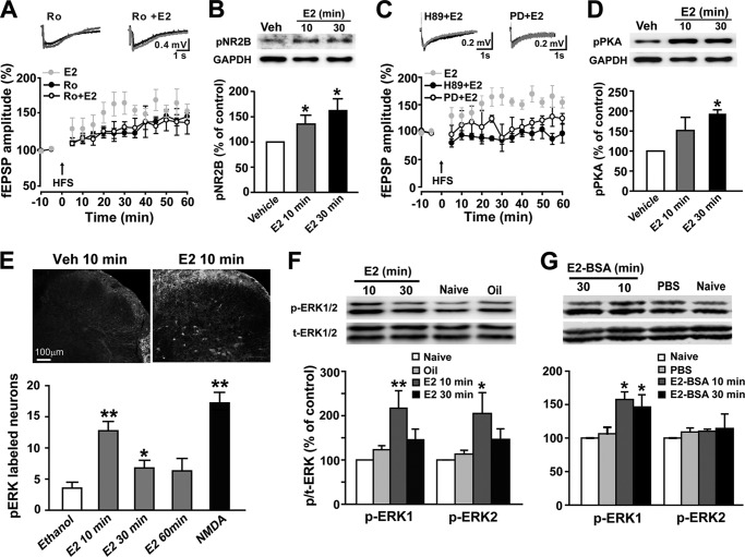
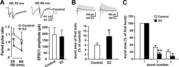
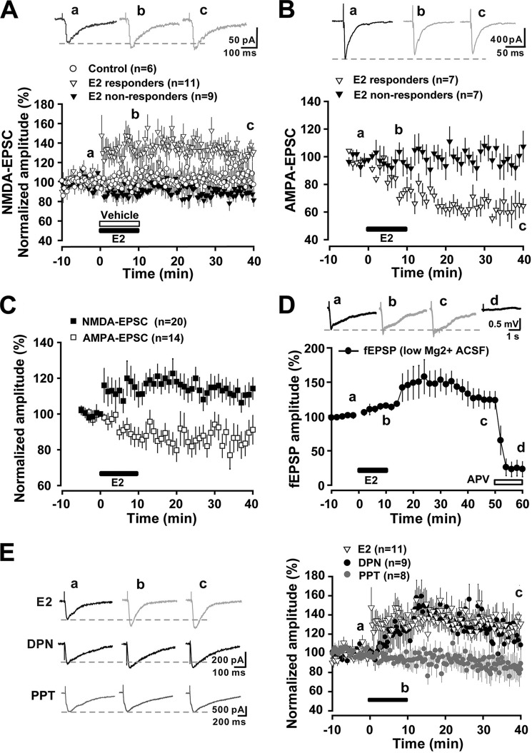
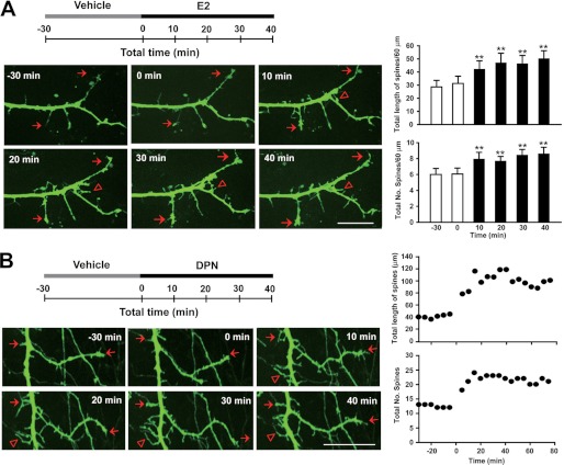
Recent evidence suggests that estrogen is synthesized in the spinal dorsal horn and plays a role in nociceptive processes. However, the cellular and molecular mechanisms underlying these effects remain unclear. Using electrophysiological, biochemical, and morphological techniques, we here demonstrate that 17β-estradiol (E2), a major form of estrogen, can directly modulate spinal cord synaptic transmission by 1) enhancing NMDA receptor-mediated synaptic transmission in dorsal horn neurons, 2) increasing glutamate release from primary afferent terminals, 3) increasing dendritic spine density in cultured spinal cord dorsal horn neurons, and 4) potentiating spinal cord long term potentiation (LTP) evoked by high frequency stimulation (HFS) of Lissauer's tract. Notably, E2-BSA, a ligand that acts only on membrane estrogen receptors, can mimic E2-induced facilitation of HFS-LTP, suggesting a nongenomic action of this neurosteroid. Consistently, cell surface biotinylation demonstrated that three types of ERs (ERα, ERβ, and GPER1) are localized on the plasma membrane of dorsal horn neurons. Furthermore, the ERα and ERβ antagonist ICI 182,780 completely abrogates the E2-induced facilitation of LTP. ERβ (but not ERα) activation can recapitulate E2-induced persistent increases in synaptic transmission (NMDA-dependent) and dendritic spine density, indicating a critical role of ERβ in spinal synaptic plasticity. E2 also increases the phosphorylation of ERK, PKA, and NR2B, and spinal HFS-LTP is prevented by blockade of PKA, ERK, or NR2B activation. Finally, HFS increases E2 release in spinal cord slices, which can be prevented by aromatase inhibitor androstatrienedione, suggesting activity-dependent local synthesis and release of endogenous E2.


Figures

FIGURE 1.
FIGURE 1.
Estrogen rapidly potentiates NMDA currents via G protein-coupled receptors in superficial dorsal horn neurons. NMDA and AMPA currents were recorded in lamina I and II neurons of spinal cord slices. E2 (100 nm to 10 μm) was bath-applied for 3–5 min. A, infusion of E2 at concentrations of 100 nm (n = 8), 1 μm (n = 7), and 10 μm (n = 12) caused a rapid, dose-dependent increase in NMDA current amplitude (infusion period indicated by bar). Top, NMDA current traces collected as controls and during E2 infusion. *, p < 0.05; **, p < 0.01 versus control. B, NMDA currents in neurons of control and E2 pretreatment slices. NMDA currents obtained from slices pretreated for 30 min with E2 (n = 11) were significantly higher than those from naive slices (n = 16). Top, NMDA current traces collected as control and pretreatment by E2. **, p < 0.01 versus naive control. C, E2 at concentrations of 1 μm (n = 11) and 10 μm (n = 17) had no effects on AMPA currents. Top, AMPA current traces collected as controls and during E2 infusion. D, loading neurons with 1 mm GDPβS abolished the acute action of E2 on NMDA currents (n = 9). E, E2 increases the NMDA/AMPA ratio. Left, the effects of E2 on the NMDA/AMPA ratio in individual cells (n = 11). Measurements were counted at the peak point of the traces at −70 mV for AMPA-EPSCs and at 40 ms after the stimulation artifacts for the traces at +40 mV for NMDA-EPSCs. Right, the effects of E2 on NMDA-EPSCs and AMPA-EPSCs. Top, typical NMDA-EPSCs recorded at +40 mV and AMPA-EPSCs at −70 mV before and after E2 application. **, p < 0.01 versus control. F, two examples showing the effects of E2 on expression of GluN1 and GluA1 in surface biotinylation-isolated membrane protein and whole cell protein extracts from cultured dorsal horn neurons and spinal slices. Neurons or slices were treated with vehicle or E2 (10 μm) for 10 min. Transferrin receptor (TrR), a marker for membrane proteins, served as a loading control. Error bars, S.E.
FIGURE 2.
FIGURE 2.
Distribution of estrogen receptors in spinal dorsal horn neurons. A, immunocytochemistry for E2-BSA-FITC and double immunofluorescence for E2-BSA-FITC and GluN1 or GluA1 in cultured spinal dorsal horn neurons. B, double immunofluorescence showed colocalization of ERα, ERβ, and GPER1 with the neuronal marker NeuN and MAP-2 in cultured dorsal horn neurons. C, double immunofluorescence showed colocalization of ERα, ERβ, and GPER1 with the neuronal marker NeuN in the spinal dorsal horn in situ. D, evidence for mERs in cultured dorsal horn neurons (a) and spinal slices (b). Surface biotinylation showed that ERα, ERβ, and GPER1 were present in the membrane protein extract. Transferrin receptor (TrR), a marker of membrane proteins, served as a loading control.
FIGURE 3.
FIGURE 3.
The role of E2 in spinal LTP of fEPSPs. A, three trains of HFS reliably induced LTP of fEPSPs in the spinal dorsal horn (n = 7). E2 (10 μm) raised the amplitude of LTP (n = 5). E2-BSA (10 μm) induced an analogous enhancement of LTP magnitude (n = 5). E2-induced enhancement of LTP was blocked by 1 μm ICI 182,780 (n = 7). Top, traces collected during base-line recording (black line) and 60 min after three trains of HFS (gray lines) in different groups. B, pretreatment with APV (50 μm) completely impaired three-train HFS-induced LTP of fEPSPs (n = 5). Top, representative fEPSPs recorded at the times indicated by the letters. C and D, neither E2 (10 μm) nor E2-BSA (10 μm) affected basal fEPSPs in normal ACSF (n = 4–6). Top panels, representative fEPSPs recorded at the times indicated by the letters. E, one-train HFS (arrows) produced more potentiation following preinfusion with E2 (n = 12) compared with control slices (n = 13). Top, traces collected from slices during base-line recording (black line) and 30 min after delivery of one train of HFS (gray lines). F, E2 levels in the perfusate of spinal slices before and after HFS. E2 levels were significantly elevated at 5 and 10 min after HFS compared with conditioning stimulation. Preincubation of slices in ACSF containing androstatrienedione (ATD 20 and 100 μm) for 30 min significantly attenuated the HFS-induced increase in E2 concentrations. *, p < 0.05; **, p < 0.01 versus controls; $, p < 0.05 versus HFS. Error bars, S.E.
FIGURE 4.
FIGURE 4.
Involvement of NR2B, PKA, and ERK activation in E2-induced facilitation of spinal LTP. A, at a dose of 0.3 μm, Ro 25-6981 (Ro) eliminated E2-induced potentiation of HFS-LTP (n = 8) but did not block HFS-LTP of fEPSPs in the spinal dorsal horn (n = 7). Top, representative fEPSPs recorded before HFS (black lines) and 60 min after HFS (gray lines). B, Western blot analysis revealed rapid phosphorylation of NR2B by E2 exposure in cultured spinal dorsal horn neurons (n = 4). Top, representative Western blot for pNR2B protein from vehicle- and E2-treated neurons. C, pretreatment with either H89 (1 μm) (n = 8) or PD 98059 (PD; 50 μm) (n = 4) eliminated E2-induced LTP enhancement. D, Western blot analysis showed rapid phosphorylation of PKA by E2 exposure in cultured spinal dorsal horn neurons (n = 5). E, immunohistochemistry for pERK from spinal cord slices. Following E2 or NMDA treatment, the numbers of pERK-positive cells in the superficial layers were higher than those of vehicle controls. *, p < 0.05; **, p < 0.01 versus vehicle (0.1% ethanol) controls. F and G, intrathecal injection of E2 (75 nmol) or E2-BSA (75 nmol) significantly increased expression levels of pERK in the spinal cord dorsal horn (n = 4). Top, representative Western blot for pERK expression in naive and vehicle- and E2- (or E2-BSA)-treated rats. *, p < 0.05 versus vehicle (0.1% ethanol or 0.01 m PBS). Error bars, S.E.
FIGURE 5.
FIGURE 5.
Effects of E2 on presynaptic release probability in dorsal horn neurons. A, effects of E2 (10 μm) on the PPR. The PPR was measurably reduced at 35- and 60-ms intervals by E2 (n = 11). Top, representative traces collected as controls (black line) and after E2 application (gray line) at 30-ms (left) and 60-ms intervals (right). B, comparison of burst area during the first train of HFS relative to the area of base-line NMDA-EPSCs averaged across control slices (n = 15) and E2 pretreatment slices (n = 12). C, comparison of the extent to which the burst responses were facilitated within three trains of HFS between the two groups. The areas of burst responses 2 and 3 were estimated by expressing them as fractions of the size of the first burst in the train. The percentage increases in burst response areas across the second and third bursts in control slices were both larger than those of E2-treated slices. *, p < 0.05; **, p < 0.01 versus controls. Error bars, S.E.
FIGURE 6.
FIGURE 6.
Estrogen induces chemical LTP of NMDA-EPSCs and LTD of AMPA-EPSCs. A, E2 (10 μm) induced a chemical LTP of NMDA-EPSC in 11 of 20 neurons (responders), whereas NMDA-EPSCs remained unaltered in the remaining neurons (non-responders) as compared with vehicle-treated (ethanol) neurons (n = 6). Each point represents an averaged trace of three sweeps. Top, traces collected as controls, 5 min after E2 infusion, and after 30 min of washout. B, E2 induced a chemical LTD of AMPA-EPSC in 7 of 14 neurons (responders), whereas AMPA-EPSCs were unchanged in the remaining neurons (non-responders). C, when all E2 responders and non-responders were pooled together, a persistent potentiation of NMDA-EPSCs and a long lasting depression of AMPA-EPSCs due to E2 were revealed. D, E2 rapidly increased LT stimulation-induced (0.1 ms, 0.5–0.7 mA, 2-min interval) fEPSPs (LT-fEPSPs) in the superficial dorsal horn, and this effect was eliminated by APV (50 μm). Top, traces collected as controls, 10 min after E2 infusion, 30 min after E2 washout, and 10 min after APV (50 mm) infusion. LT-fEPSPs were recorded in low Mg2+ high Ca2+ ACSF. E, comparison of the effects of E2 (n = 11), DPN (5 μm) (n = 9), and PPT (2 μm) (n = 8) on NMDA-EPSCs. Left, traces collected as controls, 10 min after E2, DPN, or PPT infusion and after 30-min washout. Error bars, S.E.
FIGURE 7.
FIGURE 7.
Estrogen rapidly modulates dendritic spine morphogenesis. A, E2 enhances dendritic spine density and length in GFP-expressing spinal dorsal horn neurons. Top, schematic of the experiment, which involved 40-min exposure to E2 before 30-min vehicle treatment. Left, time lapse imaging of a typical neuron expressing GFP. The neuron was imaged for the 30-min vehicle treatment and then at 0, 10, 20, 30, and 40 min after treatment with E2. Triangles indicate novel spines; arrows represent persistent spines. Right, the total length and number of spines were rapidly increased by E2 at all time points observed. **, p < 0.01 versus before E2. B, an example showing the time course of DPN-induced increases in spine length and spine number. Scale bars, 20 μm. Error bars, S.E.

References

    1. Maggiolini M., Picard D. (2010) The unfolding stories of GPR30, a new membrane-bound estrogen receptor. J. Endocrinol. 204, 105–114 - PubMed
    1. Peng H. Y., Chen G. D., Tung K. C., Chien Y. W., Lai C. Y., Hsieh M. C., Chiu C. H., Lai C. H., Lee S. D., Lin T. B. (2009) Estrogen-dependent facilitation on spinal reflex potentiation involves the Cdk5/ERK1/2/NR2B cascade in anesthetized rats. Am. J. Physiol. Endocrinol. Metab. 297, E416–E426 - PubMed
    1. Tang B., Ji Y., Traub R. J. (2008) Estrogen alters spinal NMDA receptor activity via a PKA signaling pathway in a visceral pain model in the rat. Pain 137, 540–549 - PMC - PubMed
    1. Liu N. J., Chakrabarti S., Schnell S., Wessendorf M., Gintzler A. R. (2011) Spinal synthesis of estrogen and concomitant signaling by membrane estrogen receptors regulate spinal κ- and μ-opioid receptor heterodimerization and female-specific spinal morphine antinociception. J. Neurosci. 31, 11836–11845 - PMC - PubMed
    1. Evrard H. C., Balthazart J. (2004) Rapid regulation of pain by estrogens synthesized in spinal dorsal horn neurons. J. Neurosci. 24, 7225–7229 - PMC - PubMed

Publication types