. 2012 Sep;53(5):1028-35.
doi: 10.3349/ymj.2012.53.5.1028.

Development of the rectus abdominis and its sheath in the human fetus

Affiliations

Development of the rectus abdominis and its sheath in the human fetus

Jae Do Yang et al. Yonsei Med J. 2012 Sep.

Abstract

Purpose: Although the rectus abdominis and its sheath are well known structures, their development in the human fetus is poorly understood.

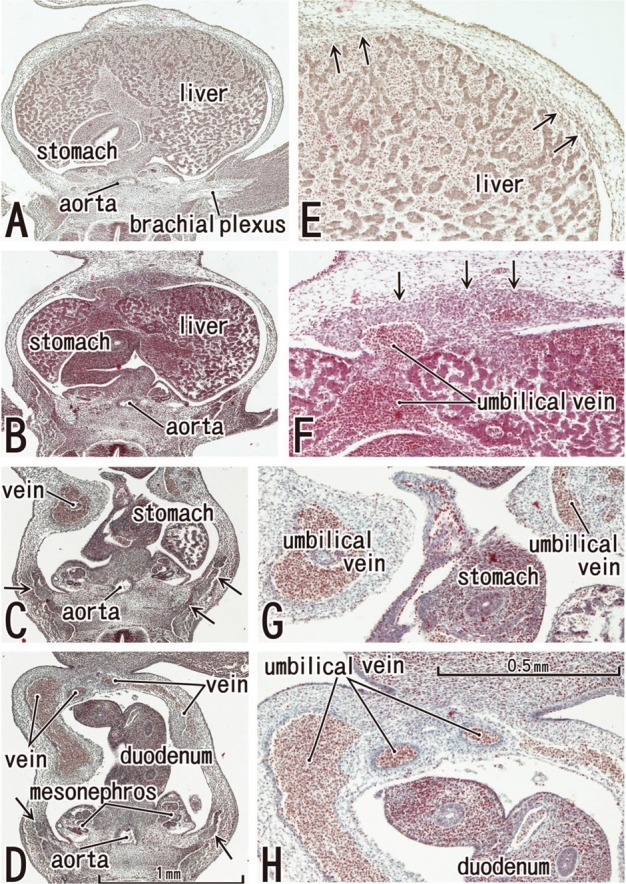
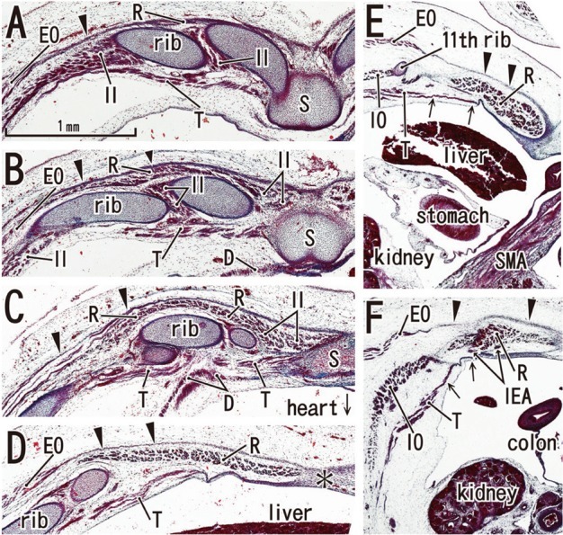
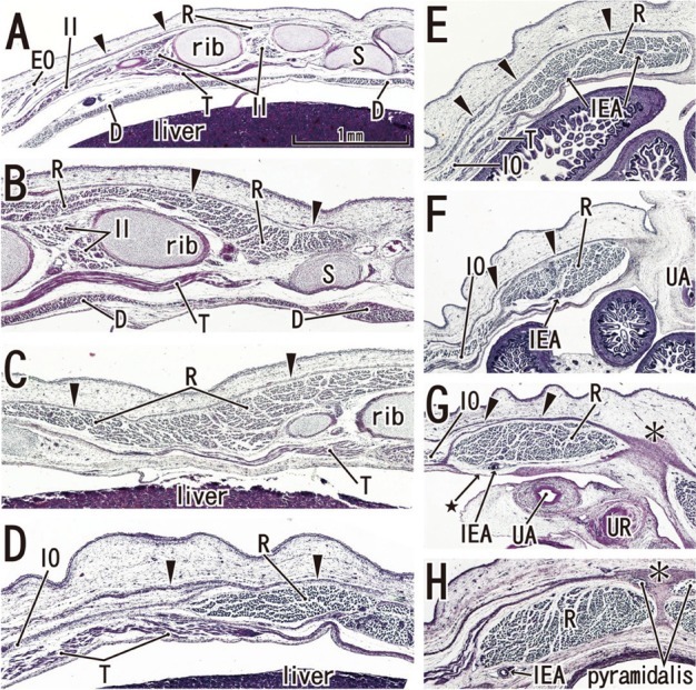
Materials and methods: We examined rectus abdominis and sheath development in semiserial horizontal sections of 18 fetuses at 5-9 weeks of gestation.

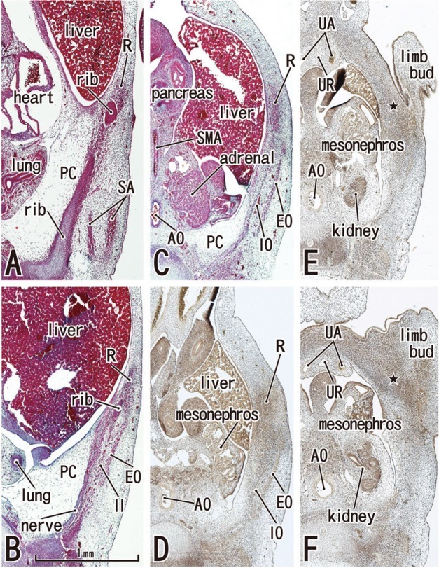
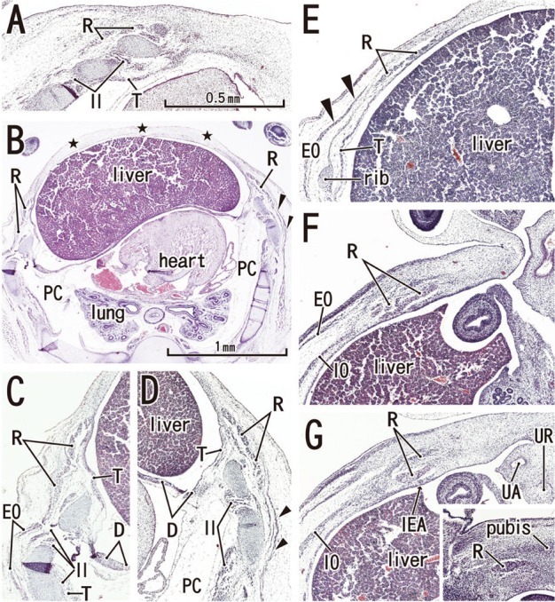
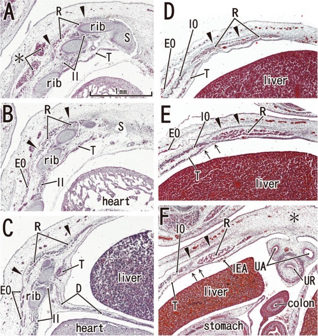
Results: Rectus muscle differentiation was found to commence above the umbilicus at 6 weeks and extend inferiorly. Until closure of the anterior chest wall via fusion of the bilateral sternal anlagen (at 7 weeks), the anterior rectal sheath originated from the external oblique and developed towards the medial margin of the rectus abdominis at all levels, including the supracostal part. After formation of the anterior sheath, fascial laminae from the internal oblique and transversus abdominis contributed to formation of the posterior rectus sheath. However, the posterior sheath was absent along the supracostal part of the rectus abdominis, as the transversus muscle fibers reached the sternum or the midline area. Therefore, it appeared that resolution of the physiological umbilical hernia (8-9 weeks) as well as chest wall closure was not required for development of the rectus abdominis and its sheath. Conversely, in the inferior part of the two largest fetal specimens, after resolution of the hernia, the posterior sheath underwent secondary disappearance, possibly due to changes in mechanical stress.

Conclusion: Upward extension of the rectus abdominis suddenly stopped at the margin of the inferiorly developing pectoralis major without facing the external intercostalis. The rectus thoracis, if present, might correspond to the pectoralis.


Conflict of interest statement

The authors have no financial conflicts of interest.

Figures

Fig. 1
Fig. 1
The abdominal wall is occupied by the umbilical veins in tilted horizontal sections of a 5-week embryo (CRL 7 mm). HE staining. Panel A (or Panel D) is the most superior (or inferior) in the figure. Panels E-H are higher magnification views of the primitive rectus abdominis shown in panels A-D, respectively. Panel C displays the level of the umbilical hernia. Arrows in panels B-D (or E and F) indicate developing ribs (or candidate anlagen of the abdominal wall muscles). The plate-like, midline condensation (arrows in panel F) appears to correspond to the abdominal band in mice and rats (see text for explanation). Panels A-D or panels E-H were prepared at the same magnification (scale bar in panels D and H).
Fig. 2
Fig. 2
Abdominal wall muscles appear in horizontal sections of a 6-week embryo (CRL 16 mm). HE staining (panels A-C) and silver impregnation (panels D-F). Panel A (or Panel F) is the most superior (or inferior) level of the figure. The rectus abdominis (R) appears to be differentiated at levels above the umbilicus (panels A and B), but it is identified as a mesenchymal cell cluster alongside the umbilicus (panels C and D) and is difficult to identify below the umbilicus (star in panels E and F). Likewise, the oblique muscles (EO, IO) are not identified below the umbilicus (panels E and F). The rectus abdominis is located in the lateral part of the body wall rather than the anterior site. The rectus sheath (arrowheads) was not seen at any level. The diaphragm has not yet developed. The future inferior part of the pleural cavity is filled with loose tissue (PC in panels A-C). All panels were prepared at the same magnification (scale bar in panel B). In all figures including Fig. 1, the upper side of each panel corresponds to the anterior or ventral side of the body. AO, aorta; SA, serratus anterior; SMA, superior mesenteric artery; UA, umbilical artery; UR, urachus; EO, external oblique; IO, internal oblique; PC, primitive pleural cavity.
Fig. 3
Fig. 3
Anterior rectal sheath appears in horizontal sections of a 7-week embryo (CRL 21 mm). HE staining. Panel A (or Panel G) is the most superior (or inferior) level of the figure. The rectus abdominis extends superiorly in the superficial side of the rib cage (panels A-D). However, the anterior chest wall is not closed by the sternum but by the surface ectoderm and underlining mesenchymal tissues (stars in panel B). Panels C and D are higher magnification views of the bilateral rectus muscles shown in panel B. The PC is filled with loose mesenchymal tissue (panels B and C). Panel E, the level of the umbilical vein exiting the liver, with the ribs also present. The anterior sheath (arrowheads), originating from the external oblique (EO), covers the rectus abdominis at levels above the umbilicus (panels B-E). In panel F (the level of the umbilical hernia) and panel G (a level below the umbilicus), the rectus sheath as well as the transversus (T) is difficult to identify. In panel G, the rectus abdominis is artifactually divided into two parts during the histological procedure. The rectus abdominis is reduced in size at the level below the umbilicus (panel G). An insert of panel G demonstrates the most inferior part of the muscle comprising of a single small band adjacent to the pubic bone anlage (pubis). Panels A and C-G were prepared at the same magnification (scale bar in panel A). D, diaphragm; IEA, inferior epigastric artery; IO, internal oblique; PC, primitive pleural cavity; T, transverses abdominis/thoracis; UA, umbilical artery; UR, urachus.
Fig. 4
Fig. 4
Posterior rectus sheath appears in horizontal sections of an 8-week embryo (CRL 29 mm). HE staining. Panel A (or Panel F) is the most superior (or inferior) level of the figure. In panels A-C, the rectus abdominis (R) attaches to the anterior aspect of the ribs, but it does not extend medially to reach the sternum (S). The rectus abdominis is located at the superficial side of the internal intercostal (II). The asterisk in panel A indicates bleeding. The anterior sheath (arrowheads), originating from the external oblique (EO), covers the rectus abdominis at all levels (panels A-F). In panel D, the level of the umbilical vein exiting the liver, note the absence of ribs. In panel E (a level of the umbilical hernia) and panel F (a level below the umbilicus), the posterior sheath (arrows) originates from the transversus abdominis (T). In panel F, the midline area is composed of loose mesenchymal tissue (asterisk) and the anterior sheath (arrowheads) does not reach the midline area. All panels were prepared at the same magnification (scale bar in panel A). D, diaphragm; IEA, inferior epigastric artery; IO, internal oblique; T, transverses abdominis/thoracis; UA, umbilical artery; UR, urachus.
Fig. 5
Fig. 5
Posterior rectus sheath becomes clear in horizontal sections of a 9-week fetus (CRL 35 mm). HE staining. Panel A (or Panel F) is the most superior (or inferior) level of the figure. The bilateral sternal anlagen have already fused (panels A and B). The supracostal part of the rectus abdominis (R) is located on the superficial side of (panel A), but in panels B and C, at the same layer as the internal intercostal (II). The anterior sheath (arrowheads), originating from the external oblique (EO), covers the rectus abdominis at all levels (panels A-F). In panel D, the level of the umbilical vein exiting the liver, the ribs are seen. A midline raphe-like structure can be seen (asterisk in panel D). Panel E, a level of the umbilical hernia, which includes the 11th rib that receives the internal oblique (IO). In panel F, a level below the umbilicus, three lateral muscles are separated by relatively wide loose mesenchymal tissues. Arrows in panels E and F indicate the posterior sheath originating from the transversus abdominis (T). All panels were prepared at the same magnification (scale bar in panel A). SMA, superior mesenteric artery. D, diaphragm; IEA, inferior epigastric artery; S, sternum.
Fig. 6
Fig. 6
Secondary change of the posterior rectus sheath after resolution of the umbilical hernia: horizontal sections of a 9-week fetus (CRL 36 mm). HE staining. Panel A (or Panel H) is the most superior (or inferior) level of the figure. In panels A-C, the rectus abdominis (R) is well developed in the anterior aspect of the internal intercostal (II). In panel D, the level of the umbilical vein exit, note the absence of ribs. The anterior sheath (arrowheads) is clearly identified at the medial side of the rectus abdominis (panels E-H). At a level below the umbilicus, part of the posterior sheath is absent (arrow with star in panel G) and the midline area (asterisk in panels G and H) is composed of dense connective tissue. The inferior epigastric artery (IEA) runs upward from panel H to panel F as a single trunk, and divides into two major branches at a level between panels E and F. All panels were prepared at the same magnification (scale bar in panel A). D, diaphragm; IO, internal oblique; T, transverses abdominis/thoracis; UA, umbilical artery; UR, urachus; EO, external oblique; S, sternum.

References

    1. Williams PL. Gray's Anatomy. 38th ed. Edinburgh: Churchill Livingstone; 1995. pp. 825–829.
    1. Munger GT, Munger BL. Differentiation of the anterior body wall and truncal epidermis and associated co-migration of cutaneous nerves and mesenchyme. Anat Rec. 1991;231:261–274. - PubMed
    1. Gościcka D, Murawski E. Tendinous intersections of the rectus abdominis muscle in human fetuses. Folia Morphol (Warsz) 1980;39:427–434. - PubMed
    1. Niikura H, Okamoto S, Nagase S, Takano T, Murakami G, Tatsumi H, et al. Fetal development of the human gubernaculum with special reference to the fasciae and muscles around it. Clin Anat. 2008;21:547–557. - PubMed
    1. Rizk NN. A new description of the anterior abdominal wall. Anat Rec. 1976;184:515. - PMC - PubMed

Publication types