. 2012 Nov;11(11):1230-44.
doi: 10.1074/mcp.M112.020594. Epub 2012 Aug 6.

Interactome-wide analysis identifies end-binding protein 1 as a crucial component for the speck-like particle formation of activated absence in melanoma 2 (AIM2) inflammasomes

Affiliations

Interactome-wide analysis identifies end-binding protein 1 as a crucial component for the speck-like particle formation of activated absence in melanoma 2 (AIM2) inflammasomes

Li-Jie Wang et al. Mol Cell Proteomics. 2012 Nov.

Abstract

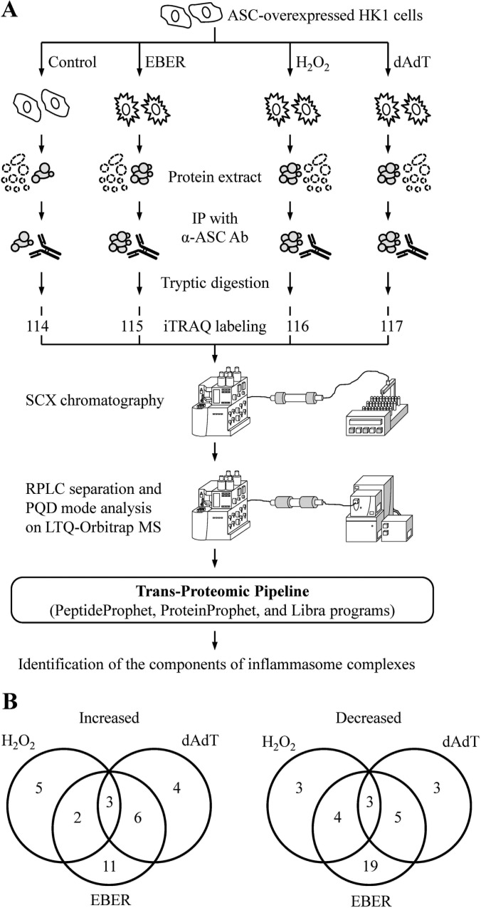
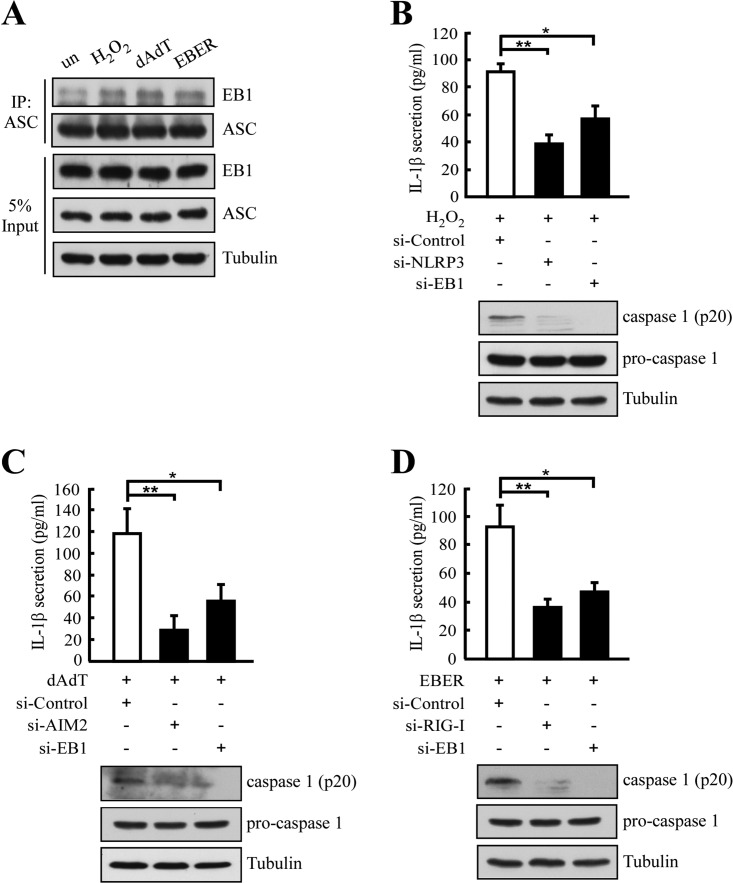
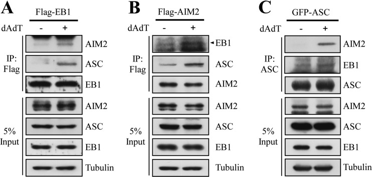
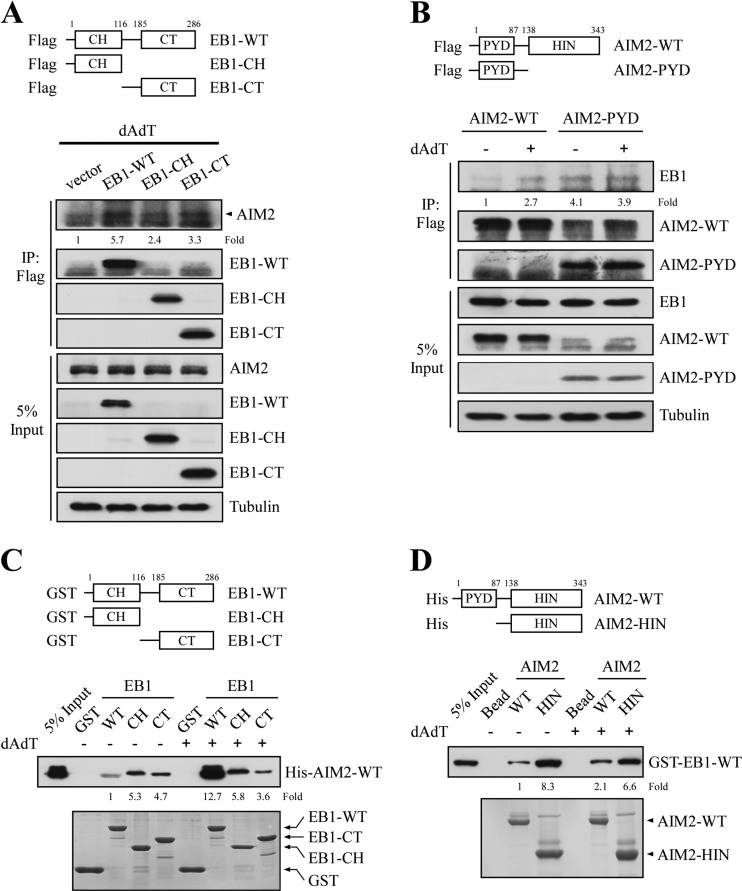
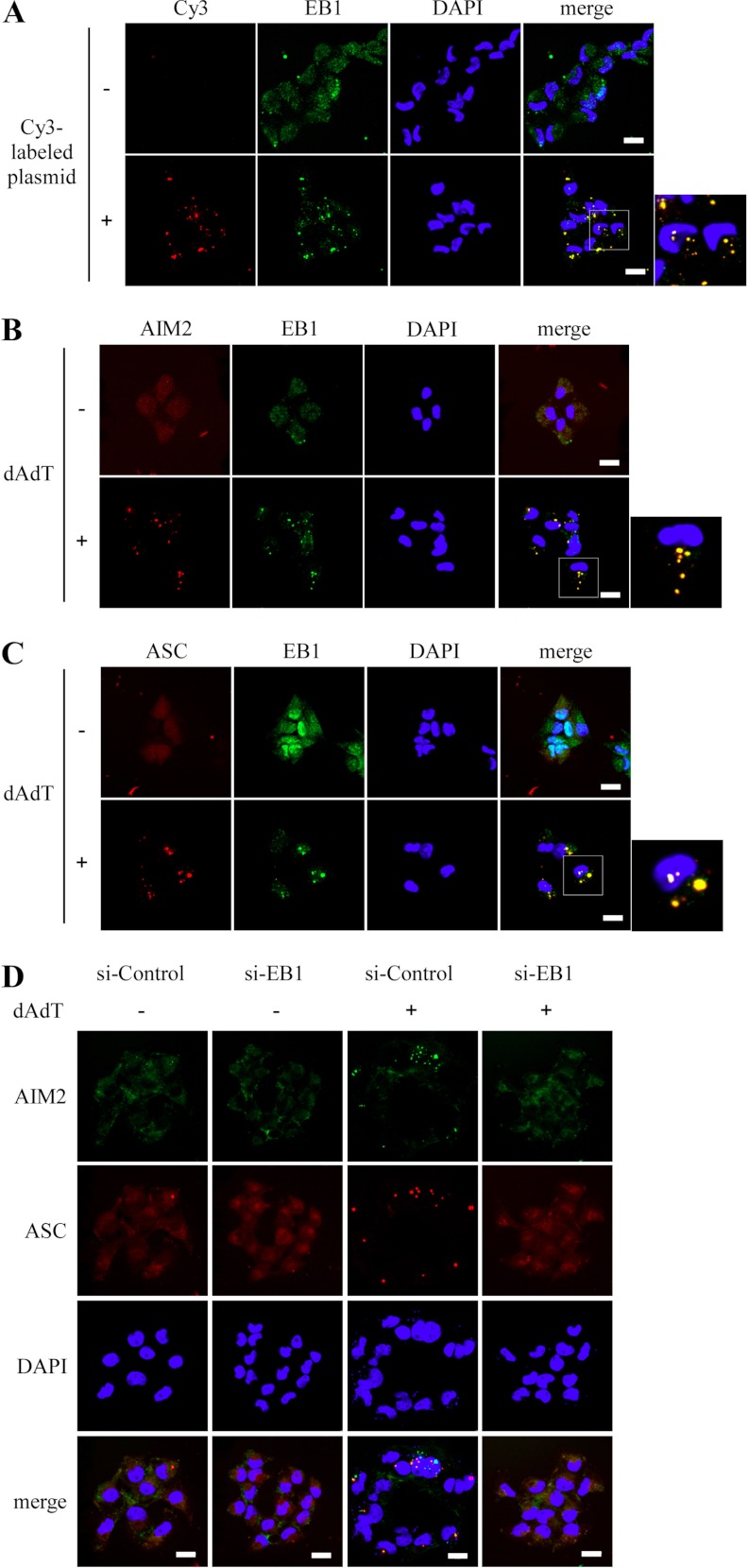
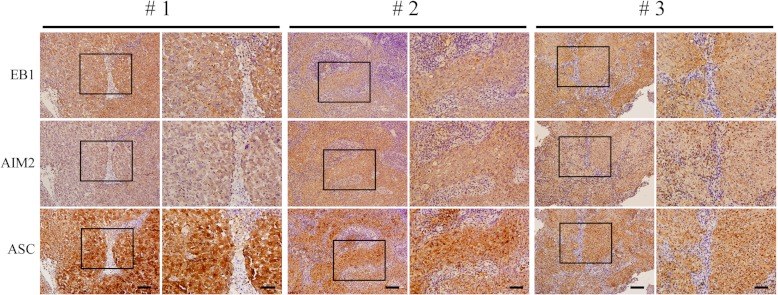
Inflammasomes are cytoplasmic receptors that can recognize intracellular pathogens or danger signals and are critical for interleukin 1β production. Although several key components of inflammasome activation have been identified, there has not been a systematic analysis of the protein components found in the stimulated complex. In this study, we used the isobaric tags for relative and absolute quantification approach to systemically analyze the interactomes of the NLRP3, AIM2, and RIG-I inflammasomes in nasopharyngeal carcinoma cells treated with specific stimuli of these interactomes (H2O2, poly (dA:dT), and EBV noncoding RNA, respectively). We identified a number of proteins that appeared to be involved in the interactomes and also could be precipitated with anti-apoptosis-associated speck-like protein containing caspase activation and recruitment domain antibodies after stimulation. Among them, end binding protein 1 was an interacting component in all three interactomes. Silencing of end binding protein 1 expression by small interfering RNA inhibited the activation of the three inflammasomes, as indicated by reduced levels of interleukin 1β secretion. We confirmed that end binding protein 1 directly interacted with AIM2 and ASC in vitro and in vivo. Most importantly, fluorescence confocal microscopy showed that end binding protein 1 was required for formation of the speck-like particles that represent activation of the AIM2 inflammasome. In nasopharyngeal carcinoma tissues, immunohistochemical staining showed that end binding protein 1 expression was elevated and significantly correlated with AIM2 and ASC expression in nasopharyngeal carcinoma tumor cells. In sum, we profiled the interactome components of three inflammasomes and show for the first time that end binding protein 1 is crucial for the speck-like particle formation that represents activated inflammasomes.


Figures

Fig. 1.
Fig. 1.
Differentially expressed proteins in the activated inflammasome complexes of HK1/ASC cells, as assessed by iTRAQ. A, A flow chart showing our iTRAQ-based proteomic analysis of inflammasome complexes. B, The numbers of proteins found to be elevated or decreased after H2O2, poly (dA:dT) or EBER treatment, as assessed by iTRAQ.
Fig. 2.
Fig. 2.
EB1 is involved in inflammasome complexes and is required for their activity. A, Pull-down assays of inflammasome complexes from cells treated with NLRP3-, AIM2-, or RIG-I-specific stimuli. Complexes were immunoprecipitated from HK1/ASC cells using anti-ASC antibodies, and EB1 levels were detected with a specific antibody. The amount of ASC was used as the pull-down control. B, HK1 cells were pretreated with control, NLRP3, or EB1 siRNA for 48 h and then treated with H2O2 (10 μm). After 24 h, media were collected for IL-1β ELISA. C, HK1 cells were pretreated with control, AIM2, or EB1 siRNA for 48 h and treated with poly (dA:dT) for 2 h. After a further incubation for 10 h, media were collected for IL-1β ELISA. D, HK1 cells were prepared as described in (C), but treated with EBER instead of poly (dA:dT). Cell extracts were collected, and then indicated molecules were detected by specific antibodies. Symbols: *, p < 0.05 and **, p < 0.01. The results are presented as the means ± S.D. of three experiments.
Fig. 3.
Fig. 3.
EB1 associates with the AIM2 inflammasome after poly (dA:dT) treatment of NPC TW02 cells. A, NPC TW02 cells were transfected with flag-EB1 for 48 h, treated with/without poly (dA:dT) for 2 h, and then further incubated for 4 h. Cell extracts were collected and subjected to immunoprecipitation with an anti-Flag matrix, and Western blotting of the indicated proteins was performed using specific antibodies. Flag-EB1 was detected using an anti-Flag antibody. B, NPC TW02 cells were transfected with Flag-AIM2 for 48 h, treated with/without poly (dA:dT) for 2 h, and incubated for an additional 4 h. Cell extracts were collected and subjected to immunoprecipitation using an anti-Flag matrix. The indicated proteins were detected by Western blotting. Flag-AIM2 was detected using an anti-Flag antibody. C, NPC TW02 cells were transfected with GFP-ASC for 48 h, treated with/without poly (dA:dT) for 2 h, and further incubated for 4 h. Cell extracts were collected and subjected to immunoprecipitation using anti-ASC antibodies. The indicated proteins were detected by Western blotting. GFP-ASC was detected using an anti-GFP antibody.
Fig. 4.
Fig. 4.
EB1 directly interacts with AIM2 in vitro. A, NPC TW02 cells were transfected with empty vector or vectors encoding Flag-EB1-WT, Flag-EB1-CH, or Flag-EB1-CT. After 48 h, the cells were treated with poly (dA:dT) for 2 h and incubated for 4 h, and then harvested for immunoprecipitation using an anti-Flag matrix. The levels of AIM2 were assessed by Western blotting after immunoprecipitation. The fold-change numbers were obtained from three independent experiments. B, HK1 cells stably expressing Flag-AIM2-WT or Flag-AIM2-PYD (introduced by lentiviral infection) were treated with or without poly (dA:dT) and subjected to immunoprecipitation using an anti-Flag matrix. The levels of EB1 were assessed by Western blotting. C, Purified GST, GST-EB1-WT, GST-EB1-CH, and GST-EB1-CT fusion proteins were immobilized on glutathione agarose, and then separately incubated with purified His-AIM2-WT fusion proteins for 24 h. The bound proteins were washed, and then analyzed by Western blotting with an anti-His antibody. The fold-change numbers were derived from three independent experiments. The amounts of GST fusion proteins were assessed by Coomassie blue staining. D, Purified His-AIM2-WT or His-AIM2-HIN were immobilized on Ni-charged agarose, and then incubated with purified GST-EB1-WT for 24 h. The bound proteins were washed, and then analyzed by Western blotting with an anti-GST antibody. The fold-change numbers were obtained from three independent experiments. The amount of His fusion proteins were assessed by Coomassie blue staining.
Fig. 5.
Fig. 5.
EB1 colocalizes with the AIM2 inflammasome and is required for speck-like particle formation in NPC TW02 cells. A, Cells were treated with or without Cy3-labeled plasmids for 2 h, and were further incubated for 4 h. Cells were fixed and were stained with an anti-EB1 antibody, and the colocalization of Cy3-labled plasmids and EB1 was observed by confocal immunofluorescence microscopy. B, and C, Cells were transfected with DsRed-AIM2 or DsRed-ASC for 48 h, treated with poly (dA:dT) for 2 h, and further incubated for 4 h. The cells were then fixed and stained with an anti-EB1 antibody, and the colocalization of EB1 with DsRed-AIM2 or DsRed-ASC was observed by confocal immunofluorescence microscopy. D, NPC TW02 cells expressing control siRNA or EB1 siRNA were cotransfected with GFP-AIM2 and DsRed-ASC for 48 h, treated with or without poly (dA:dT) for 2 h, further incubated for 4 h, and then fixed. The colocalization of GFP-AIM2 and DsRed-ASC was observed by confocal fluorescence microscopy. Scale bar = 20 μm.
Fig. 6.
Fig. 6.
Correlation of EB1 expression with AIM2 and ASC in NPC patients. Immunohistochemical staining of EB1, AIM2, and ASC were detected by specific antibodies in consecutive NPC tissue sections from three NPC patients. The results are shown at 200× magnification (left, scale bar = 200 μm) and 400× magnification (right, scale bar = 100 μm).

References

    1. Chan A. T. (2010) Nasopharyngeal carcinoma. Ann. Oncol. 21 Suppl 7, vii308–312 - PubMed
    1. Sriamporn S., Vatanasapt V., Pisani P., Yongchaiyudha S., Rungpitarangsri V. (1992) Environmental risk factors for nasopharyngeal carcinoma: a case-control study in northeastern Thailand. Cancer Epidemiol. Biomarkers Prev. 1, 345–348 - PubMed
    1. Bei J. X., Li Y., Jia W. H., Feng B. J., Zhou G., Chen L. Z., Feng Q. S., Low H. Q., Zhang H., He F., Tai E. S., Kang T., Liu E. T., Liu J., Zeng Y. X. (2010) A genome-wide association study of nasopharyngeal carcinoma identifies three new susceptibility loci. Nat. Genet. 42, 599–603 - PubMed
    1. Chang K. P., Chang Y. T., Wu C. C., Liu Y. L., Chen M. C., Tsang N. M., Hsu C. L., Chang Y. S., Yu J. S. (2011) Multiplexed immunobead-based profiling of cytokine markers for detection of nasopharyngeal carcinoma and prognosis of patient survival. Head Neck 33, 886–897 - PubMed
    1. Apte R. N., Dotan S., Elkabets M., White M. R., Reich E., Carmi Y., Song X., Dvozkin T., Krelin Y., Voronov E. (2006) The involvement of IL-1 in tumorigenesis, tumor invasiveness, metastasis and tumor-host interactions. Cancer Metastasis Rev. 25, 387–408 - PubMed

Publication types

MeSH terms