

Corifungin, a new drug lead against Naegleria, identified from a high-throughput screen
- PMID: 22869574
- PMCID: PMC3486592
- DOI: 10.1128/AAC.00643-12
Corifungin, a new drug lead against Naegleria, identified from a high-throughput screen
Abstract
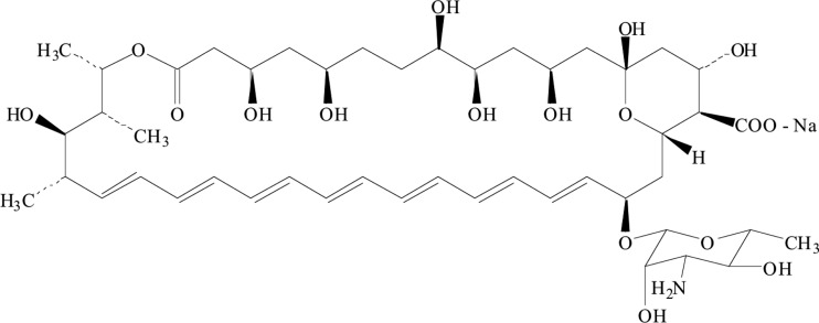
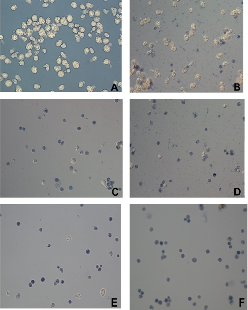
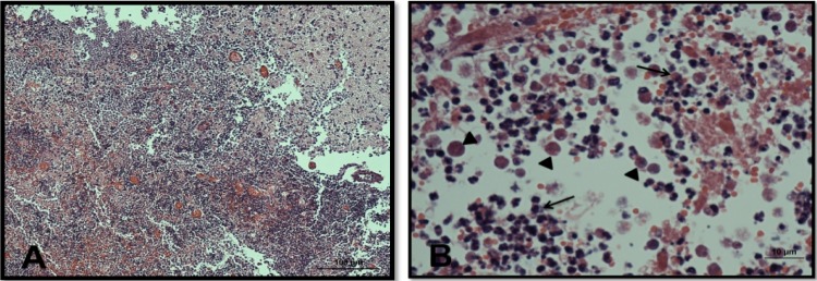
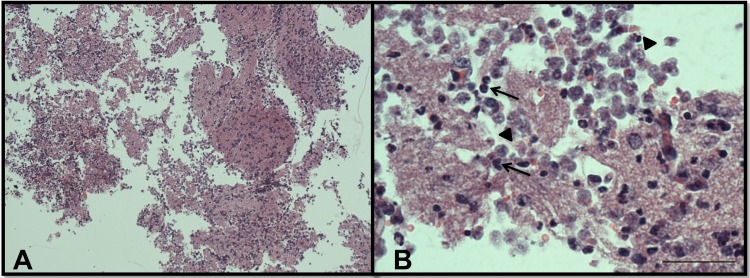
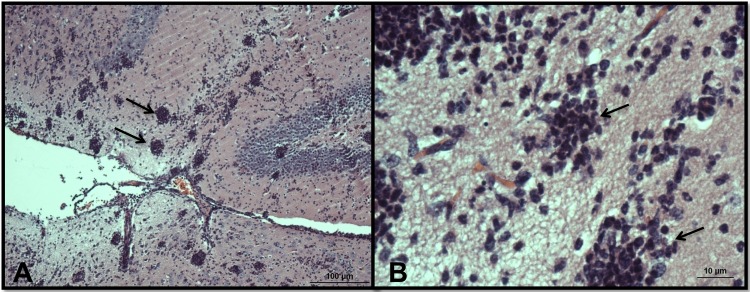
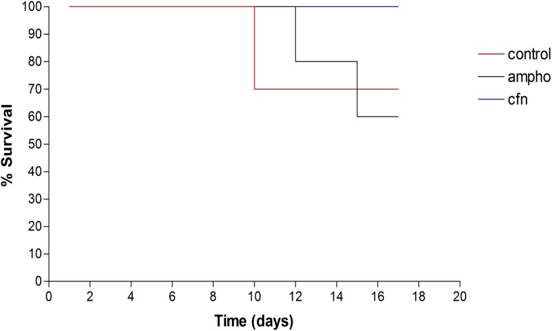
Primary amebic meningoencephalitis (PAM) is a rapidly fatal infection caused by the free-living ameba Naegleria fowleri. The drug of choice in treating PAM is the antifungal antibiotic amphotericin B, but its use is associated with severe adverse effects. Moreover, few patients treated with amphotericin B have survived PAM. Therefore, fast-acting and efficient drugs are urgently needed for the treatment of PAM. To facilitate drug screening for this pathogen, an automated, high-throughput screening methodology was developed and validated for the closely related species Naegleria gruberi. Five kinase inhibitors and an NF-kappaB inhibitor were hits identified in primary screens of three compound libraries. Most importantly for a preclinical drug discovery pipeline, we identified corifungin, a water-soluble polyene macrolide with a higher activity against Naegleria than that of amphotericin B. Transmission electron microscopy of N. fowleri trophozoites incubated with different concentrations of corifungin showed disruption of cytoplasmic and plasma membranes and alterations in mitochondria, followed by complete lysis of amebae. In vivo efficacy of corifungin in a mouse model of PAM was confirmed by an absence of detectable amebae in the brain and 100% survival of mice for 17 days postinfection for a single daily intraperitoneal dose of 9 mg/kg of body weight given for 10 days. The same dose of amphotericin B did not reduce ameba growth, and mouse survival was compromised. Based on these results, the U.S. FDA has approved orphan drug status for corifungin for the treatment of PAM.
Figures


Similar articles
-
Amphotericin B induces apoptosis-like programmed cell death in Naegleria fowleri and Naegleria gruberi.Microbiology (Reading). 2017 Jul;163(7):940-949. doi: 10.1099/mic.0.000500. Epub 2017 Jul 21. Microbiology (Reading). 2017. PMID: 28721850
-
Identification of plicamycin, TG02, panobinostat, lestaurtinib, and GDC-0084 as promising compounds for the treatment of central nervous system infections caused by the free-living amebae Naegleria, Acanthamoeba and Balamuthia.Int J Parasitol Drugs Drug Resist. 2019 Dec;11:80-94. doi: 10.1016/j.ijpddr.2019.10.003. Epub 2019 Oct 22. Int J Parasitol Drugs Drug Resist. 2019. PMID: 31707263 Free PMC article.
-
Identification of cysteine protease inhibitors as new drug leads against Naegleria fowleri.Exp Parasitol. 2018 May;188:36-41. doi: 10.1016/j.exppara.2018.03.010. Epub 2018 Mar 15. Exp Parasitol. 2018. PMID: 29551628 Free PMC article.
-
Enlightening the promising role of nanoparticle-based treatments against Naegleria fowleri-induced primary amoebic meningoencephalitis: A brain-eating disease.Microb Pathog. 2025 Feb;199:107234. doi: 10.1016/j.micpath.2024.107234. Epub 2024 Dec 17. Microb Pathog. 2025. PMID: 39701479 Review.
-
[Amebic meningoencephalitis].Brain Nerve. 2009 Feb;61(2):115-21. Brain Nerve. 2009. PMID: 19235460 Review. Japanese.
Cited by
-
Naegleria fowleri: pathogenesis, diagnosis, and treatment options.Antimicrob Agents Chemother. 2015 Nov;59(11):6677-81. doi: 10.1128/AAC.01293-15. Epub 2015 Aug 10. Antimicrob Agents Chemother. 2015. PMID: 26259797 Free PMC article. Review.
-
Differential Growth Rates and In Vitro Drug Susceptibility to Currently Used Drugs for Multiple Isolates of Naegleria fowleri.Microbiol Spectr. 2022 Feb 23;10(1):e0189921. doi: 10.1128/spectrum.01899-21. Epub 2022 Feb 9. Microbiol Spectr. 2022. PMID: 35138140 Free PMC article.
-
Phenotypic Screens Reveal Posaconazole as a Rapidly Acting Amebicidal Combination Partner for Treatment of Primary Amoebic Meningoencephalitis.J Infect Dis. 2019 Mar 15;219(7):1095-1103. doi: 10.1093/infdis/jiy622. J Infect Dis. 2019. PMID: 30358879 Free PMC article.
-
The Antifungal Drug Isavuconazole Is both Amebicidal and Cysticidal against Acanthamoeba castellanii.Antimicrob Agents Chemother. 2020 Apr 21;64(5):e02223-19. doi: 10.1128/AAC.02223-19. Print 2020 Apr 21. Antimicrob Agents Chemother. 2020. PMID: 32094126 Free PMC article.
-
Larrea tridentata: A novel source for anti-parasitic agents active against Entamoeba histolytica, Giardia lamblia and Naegleria fowleri.PLoS Negl Trop Dis. 2017 Aug 9;11(8):e0005832. doi: 10.1371/journal.pntd.0005832. eCollection 2017 Aug. PLoS Negl Trop Dis. 2017. PMID: 28793307 Free PMC article.
References
-
- Anderson K, Jamieson A. 1972. Primary amoebic meningoencephalitis. Lancet i:902–903 - PubMed
-
- Ashburn TT, Thor KB. 2004. Drug repositioning: identifying and developing new uses for existing drugs. Nat. Rev. Drug Discov. 3:673–683 - PubMed
-
- Baginski M, Sternal K, Czub J, Borowski E. 2005. Molecular modelling of membrane activity of amphotericin B, a polyene macrolide antifungal antibiotic. Acta Biochim. Pol. 52:655–658 - PubMed
-
- Berger N, Ben Bassat H, Klein BY, Laskov R. 2007. Cytotoxicity of NF-kappaB inhibitors Bay 11-7085 and caffeic acid phenethyl ester to Ramos and other human B-lymphoma cell lines. Exp. Hematol. 35:1495–1509 - PubMed
Publication types
MeSH terms
Substances
Grants and funding
LinkOut - more resources
Full Text Sources
Other Literature Sources
Miscellaneous
