Review
. 2012 Aug 6;198(3):281-93.
doi: 10.1083/jcb.201202014.

The evolving concept of cancer and metastasis stem cells

Affiliations
Review

The evolving concept of cancer and metastasis stem cells

Irène Baccelli et al. J Cell Biol. .

Abstract

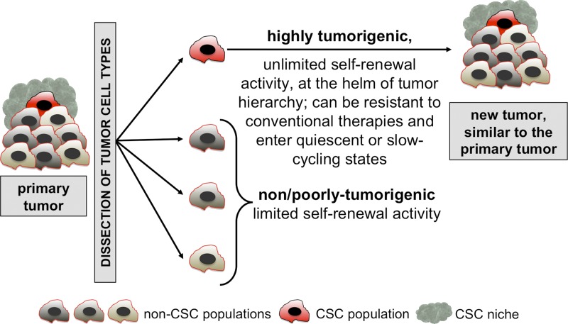
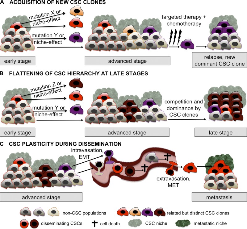
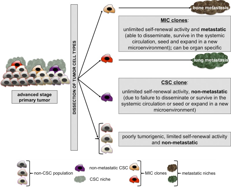
The cancer stem cell (CSC) concept, which arose more than a decade ago, proposed that tumor growth is sustained by a subpopulation of highly malignant cancerous cells. These cells, termed CSCs, comprise the top of the tumor cell hierarchy and have been isolated from many leukemias and solid tumors. Recent work has discovered that this hierarchy is embedded within a genetically heterogeneous tumor, in which various related but distinct subclones compete within the tumor mass. Thus, genetically distinct CSCs exist on top of each subclone, revealing a highly complex cellular composition of tumors. The CSC concept has therefore evolved to better model the complex and highly dynamic processes of tumorigenesis, tumor relapse, and metastasis.


Figures

Figure 1.
Figure 1.
The classical “cancer stem cell” (CSC) concept. Tumors are heterogeneous and hierarchically organized entities. Upon dissociation and transplantation into an immunocompromised animal, human CSCs can be functionally distinguished from non/poorly tumorigenic cell populations by their ability to reinitiate and grow a similar heterogeneous tumor in vivo.
Figure 2.
Figure 2.
The dynamic CSC concept. (A) Although early stage tumors might be governed by a single CSC clone, advanced stage tumors might contain several distinct but related clones, either arising from the initial CSC clone or from its differentiated progeny via mutations or via induction by the CSC niche. Targeted therapy and/or chemotherapy eliminate the tumor mass, possibly including some of the CSCs. At least one resistant CSC clone is then responsible (possibly after acquiring additional mutations) for tumor relapse. (B) Advanced stage tumors contain several distinct but related CSC clones. Some acquire enhanced self-renewal capabilities with simultaneously decreased differentiation. During tumor progression, the CSC clones compete with each other, leading to the dominance of at least one CSC clone with the subsequent loss of differentiated tumor progeny. Over time, this leads to a flattening of the hierarchical structure and to a selection of the most aggressive CSCs. Late stage tumors may thus be comprised almost exclusively of aggressive, multiresistant CSCs, a situation similar to the one proposed by the stochastic model. (C) Carcinoma CSCs display a dynamic phenotype during systemic dissemination: they are able to at least partially lose epithelial traits through EMT. It is most likely that only a subset of such disseminating CSCs are able to survive in the systemic circulation, extravasate, and reacquire epithelial features (MET) to seed in a new microenvironment and to initiate metastasis. All three scenarios illustrated in A, B, and C occur in parallel and/or during different phases of tumor progression.
Figure 3.
Figure 3.
The “metastasis initiating cell” (MIC) concept. Advanced stage primary tumors are heterogeneous and multi-hierarchical structures. Upon dissociation and transplantation in immunocompromised hosts, MICs can be functionally distinguished from CSC clones by their metastatic ability in vivo (dissemination, survival in the systemic circulation, extravasation, seeding, and expansion in a new microenvironment, termed “metastatic niche”). MICs are adapted descendants and therefore subpopulations of CSCs with metastatic capacity and, possibly, organ specificity.

References

    1. Aguirre-Ghiso J.A. 2007. Models, mechanisms and clinical evidence for cancer dormancy. Nat. Rev. Cancer. 7:834–846 10.1038/nrc2256 - DOI - PMC - PubMed
    1. Akala O.O., Park I.K., Qian D., Pihalja M., Becker M.W., Clarke M.F. 2008. Long-term haematopoietic reconstitution by Trp53-/-p16Ink4a-/-p19Arf-/- multipotent progenitors. Nature. 453:228–232 10.1038/nature06869 - DOI - PubMed
    1. Al-Hajj M., Wicha M.S., Benito-Hernandez A., Morrison S.J., Clarke M.F. 2003. Prospective identification of tumorigenic breast cancer cells. Proc. Natl. Acad. Sci. USA. 100:3983–3988 10.1073/pnas.0530291100 - DOI - PMC - PubMed
    1. American Cancer Society 2011. Cancer Facts & Figures 2011. American Cancer Society, Atlanta: 58 pp
    1. Anderson K., Lutz C., van Delft F.W., Bateman C.M., Guo Y., Colman S.M., Kempski H., Moorman A.V., Titley I., Swansbury J., et al. 2011. Genetic variegation of clonal architecture and propagating cells in leukaemia. Nature. 469:356–361 10.1038/nature09650 - DOI - PubMed

Publication types