. 2012 Aug 6;198(3):305-13.
doi: 10.1083/jcb.201204098.

Kinase-dead ATM protein causes genomic instability and early embryonic lethality in mice

Affiliations

Kinase-dead ATM protein causes genomic instability and early embryonic lethality in mice

Kenta Yamamoto et al. J Cell Biol. .

Abstract

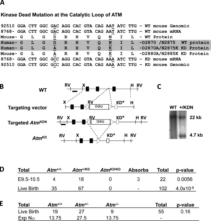
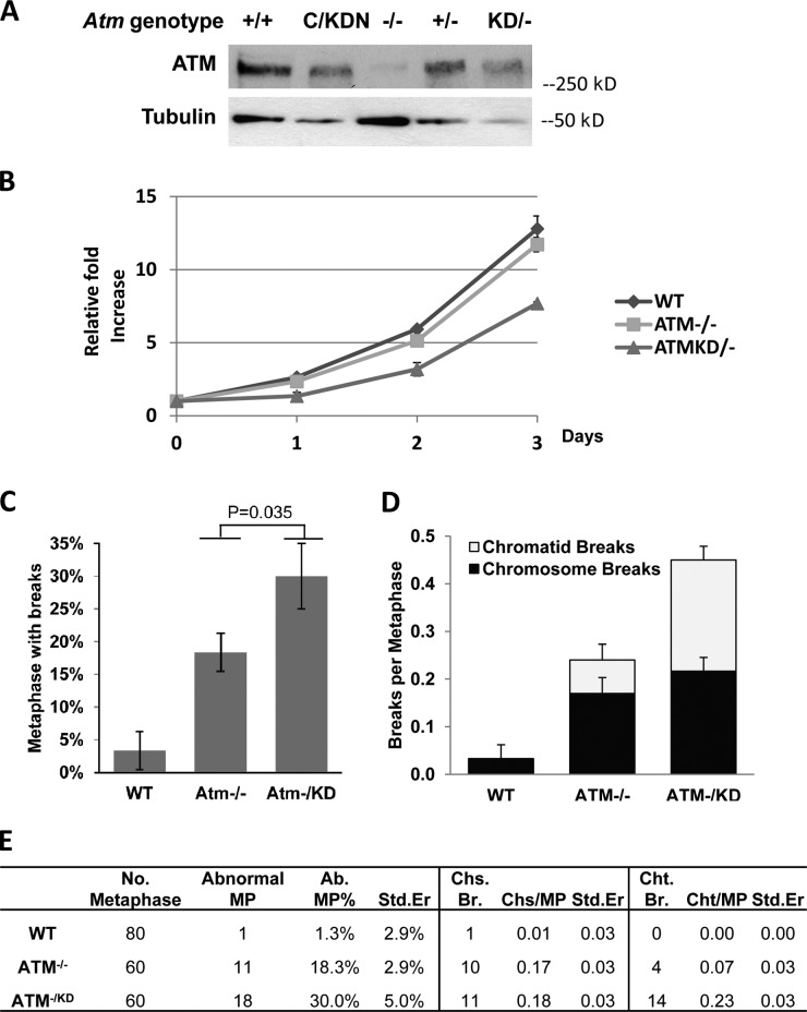
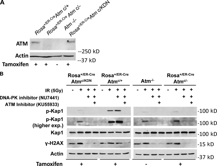
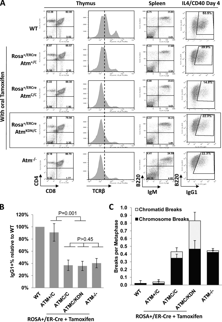
Ataxia telangiectasia (A-T) mutated (ATM) kinase orchestrates deoxyribonucleic acid (DNA) damage responses by phosphorylating numerous substrates implicated in DNA repair and cell cycle checkpoint activation. A-T patients and mouse models that express no ATM protein undergo normal embryonic development but exhibit pleiotropic DNA repair defects. In this paper, we report that mice carrying homozygous kinase-dead mutations in Atm (Atm(KD/KD)) died during early embryonic development. Atm(KD/-) cells exhibited proliferation defects and genomic instability, especially chromatid breaks, at levels higher than Atm(-/-) cells. Despite this increased genomic instability, Atm(KD/-) lymphocytes progressed through variable, diversity, and joining recombination and immunoglobulin class switch recombination, two events requiring nonhomologous end joining, at levels comparable to Atm(-/-) lymphocytes. Together, these results reveal an essential function of ATM during embryogenesis and an important function of catalytically inactive ATM protein in DNA repair.


Figures

Figure 1.
Figure 1.
Generation of the AtmKD allele. (A) The corresponding sequence of the catalytic loop of human and mouse ATM kinase are aligned, and the mutated residues are underlined. Gray shade covers the corresponding WT and mutated sequences in human ATM protein. (B) The schematic diagram represents the murine Atm locus (top), targeting vector (second row), targeted allele (AtmKDN, third row), and the neomycin (neo)-deleted mutant allele (AtmKD, bottom). The 5′ probe is marked as a black line. The exons and loxP sites are shown as solid boxes and open triangles, respectively. The exon containing the KD mutation is marked as an open box. Restriction site designations are as follows: X, XhoI; RV, EcoRV; H, HindIII. The map is not drawn to scale. (C) Southern Blot analyses of EcoRV-digested DNA from representative Atm+/+ (WT) and Atm+/KDN ES cells. (D and E) Genotype of embryos and live offspring obtained from timed intercrossing between Atm+/KD mice (D) and Atm+/− mice (E). Compared with the expected Mendelian frequency, the χ2 test p-values are 0.0056 for E9.5-–10.5 AtmKD/KD embryos and 4.01 × 10⁻⁸ for AtmKD/KD and 0.16 for Atm−/− mice at birth. Exp, expected.
Figure 2.
Figure 2.
Proliferation defects and increased genomic instability in AtmKD/− ES cells. (A) Western blot analysis of total protein (50 µg) for mouse ATM (MAT3; Sigma-Aldrich) and α-tubulin (EMD) in WT, AtmC/KDN, Atm−/−, Atm+/−, and AtmKD/− ES cells. The small amount of protein detectable in the Atm−/− lane likely arises from slight contamination of the ES cells used for this analysis with WT fibroblasts from the feeder layer. (B) Fold increase of cell number relative to day 0, as measured by modified MTT assay (Sigma-Aldrich) in WT, Atm−/−, and AtmKD/− ES cells under normal growth conditions. Three independent experiments were performed in two independently derived AtmKD/− ES cell lines. The graph and the error bars represent standard deviation derived from quadruplicated cultures in one representative experiment. (C) Frequency of metaphases with cytogenetic abnormalities in T-FISH analyses of WT, Atm+/−, and AtmKD/− ES cells. The p-value (0.035) is obtained with χ2 test. (D) The frequency of chromatid and chromosome breaks measured by T-FISH analyses in WT, Atm+/−, and AtmKD/− ES cells. The χ2 test p-value for the frequency of chromatid breaks between Atm+/− and AtmKD/− ES cells is 0.002 and for chromosome breaks is 0.101. (C and D) The error bars represent the standard deviations of multiple independent experiments. (E) Summary of cytogenetic abnormalities in WT, Atm+/−, and AtmKD/− ES cells and controls. Ab, abnormal; MP, metaphase; Chs., chromosome; Cht., chromatid; Br, break; Std.Er, standard error. The data summarize the results from three or more independent experiments using at least two independently derived cell lines of each genotype. 20 metaphases were analyzed per line per experiment.
Figure 3.
Figure 3.
ATMKD is catalytically inactive. (A) Western blot for total ATM protein in thymocytes from tamoxifen-treated Rosa+/ER-CreAtm+/C, AtmC/C, or AtmC/KDN mice and control Atm−/− mice. (B) Western blot for phosphorylated H2AX, phosphorylated KAP-1, and total KAP-1 and Actin in irradiated thymocyte (5 Gy) with or without preincubation with 15 µM ATM kinase inhibitor (KU55933; Tocris Bioscience) or 5 µM DNA-PKcs kinase inhibitor (NU7441; Tocris Bioscience). Cell lysates were collected 2 h after irradiation. Primary antibodies were used at the following dilutions: anti-Actin (1:10,000; Sigma-Aldrich), anti-ATM (1:500; MAT3; Sigma-Aldrich), anti–γ-H2AX (1:1,000; EMD Millipore), anti–KAP-1 (1:1,000; Cell Signaling Technology), antiphospho–KAP-1 (S824; 1:1,000; Bethyl Laboratories, Inc.). exp., exposure.
Figure 4.
Figure 4.
Analysis of ATM KD lymphocytes. (A) Representative flow cytometric analyses of total thymocytes stained with CD4, CD8, and TCR-β, and total splenocytes stained with B220 and IgM surface markers. For the B220/IgG1 marker staining, CD43 splenocytes were isolated and stimulated in culture with anti-CD40 and IL-4 for 4 d before staining. A total of five independent experiments were performed, and one set of representative FACS analyses is shown. The dotted line shows the median level of surface TCR-β levels in WT thymocytes. The box highlights the analyses of ATMKD/− lymphocytes. (B) Relative frequency of the IgG1+ cells among all B220+ B cells (relative to WT cells in each experiments) in Atm−/− and AtmKD/− B cells. The data represent the mean and standard deviation from at least five experiments. The χ2 test p-values are marked in the graph. (C) The frequency (per metaphase) of chromatid and chromosome breaks measured by T-FISH analyses in stimulated B cells from WT, Atm−/− mice, or tamoxifen-treated Rosa+/ER-CreAtm+/C, AtmC/C, or AtmC/KDN mice. The χ2 test p-value for the frequency of chromatid breaks between Atm+/− and AtmKD/− B cells is 0.007 and for chromosome breaks is 0.816. The table summarized the data obtained from independent experiments performed on two or three mice of each genotype (see also Fig. S3 F).

Comment in

Similar articles

Cited by

References

    1. Bakkenist C.J., Kastan M.B. 2003. DNA damage activates ATM through intermolecular autophosphorylation and dimer dissociation. Nature. 421:499–506 10.1038/nature01368 - DOI - PubMed
    1. Barlow C., Hirotsune S., Paylor R., Liyanage M., Eckhaus M., Collins F., Shiloh Y., Crawley J.N., Ried T., Tagle D., Wynshaw-Boris A. 1996. Atm-deficient mice: a paradigm of ataxia telangiectasia. Cell. 86:159–171 10.1016/S0092-8674(00)80086-0 - DOI - PubMed
    1. Barone G., Groom A., Reiman A., Srinivasan V., Byrd P.J., Taylor A.M. 2009. Modeling ATM mutant proteins from missense changes confirms retained kinase activity. Hum. Mutat. 30:1222–1230 10.1002/humu.21034 - DOI - PubMed
    1. Bhatti S., Kozlov S., Farooqi A.A., Naqi A., Lavin M., Khanna K.K. 2011. ATM protein kinase: the linchpin of cellular defenses to stress. Cell. Mol. Life Sci. 68:2977–3006 10.1007/s00018-011-0683-9 - DOI - PMC - PubMed
    1. Borghesani P.R., Alt F.W., Bottaro A., Davidson L., Aksoy S., Rathbun G.A., Roberts T.M., Swat W., Segal R.A., Gu Y. 2000. Abnormal development of Purkinje cells and lymphocytes in Atm mutant mice. Proc. Natl. Acad. Sci. USA. 97:3336–3341 10.1073/pnas.050584897 - DOI - PMC - PubMed

Publication types

MeSH terms