. 2012 Aug 21;109(34):13668-73.
doi: 10.1073/pnas.1200872109. Epub 2012 Aug 6.

A paradoxical teratogenic mechanism for retinoic acid

Affiliations

A paradoxical teratogenic mechanism for retinoic acid

Leo M Y Lee et al. Proc Natl Acad Sci U S A. .

Abstract

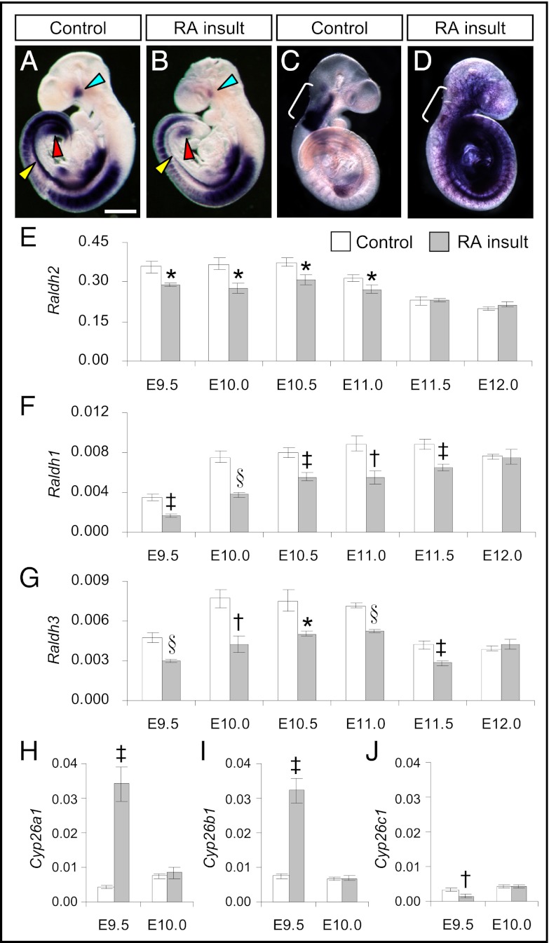
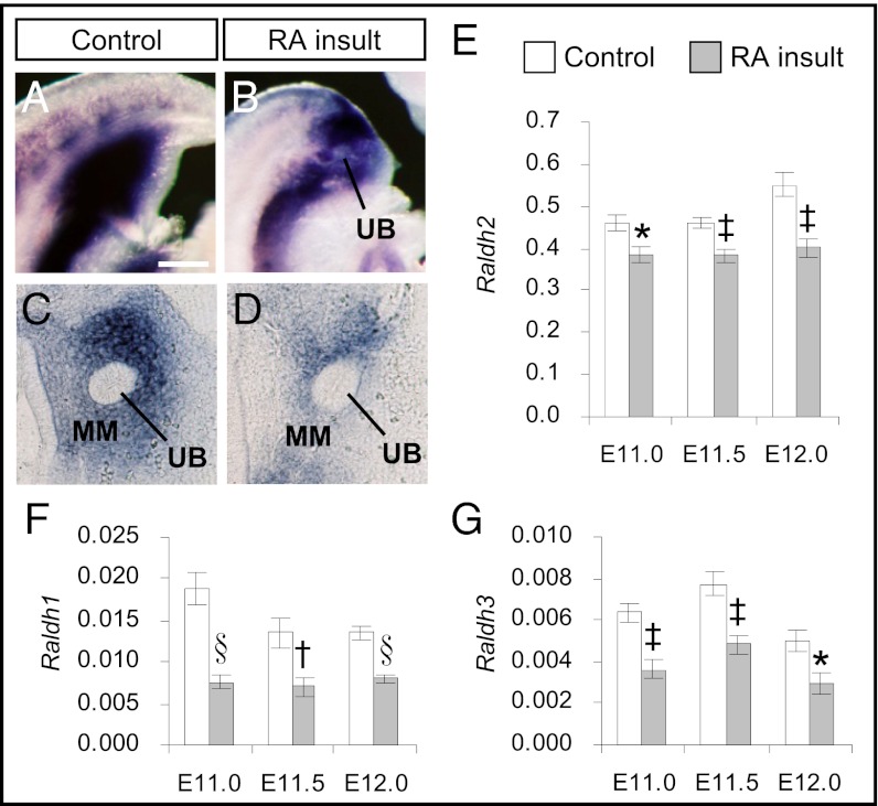
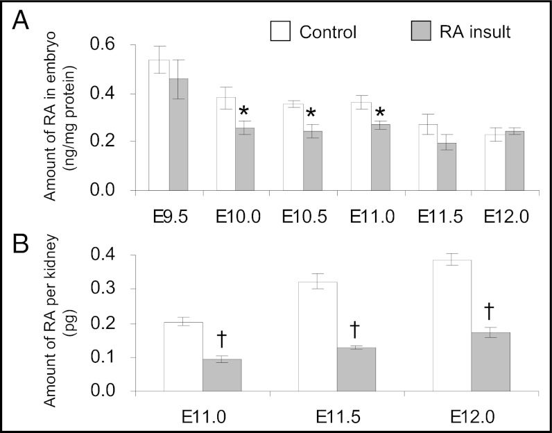
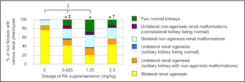
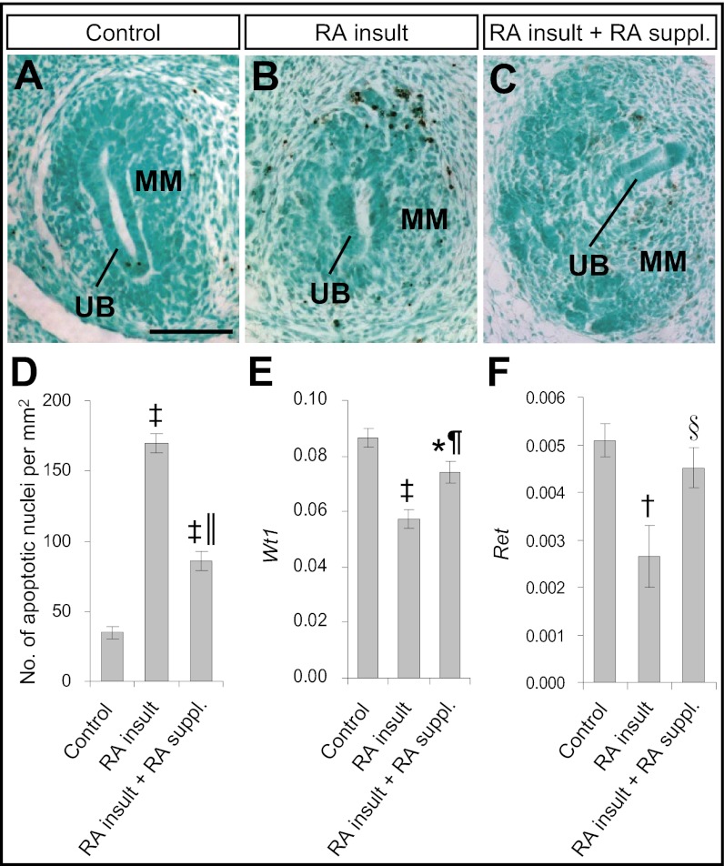
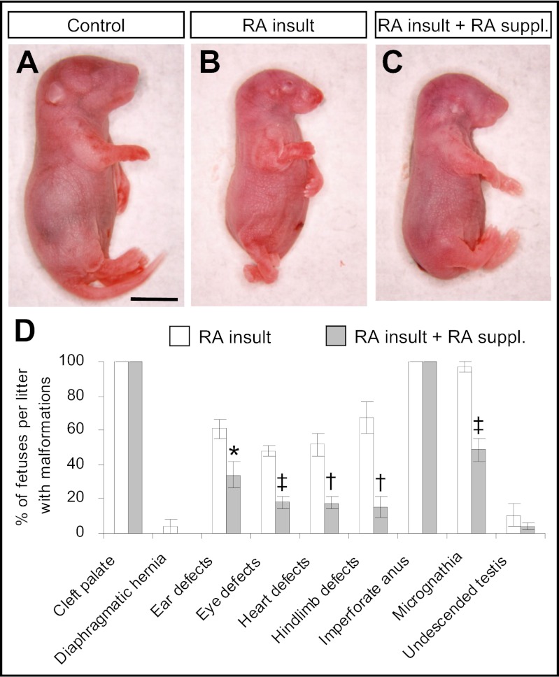
Retinoic acid, an active metabolite of vitamin A, plays essential signaling roles in mammalian embryogenesis. Nevertheless, it has long been recognized that overexposure to vitamin A or retinoic acid causes widespread teratogenesis in rodents as well as humans. Although it has a short half-life, exposure to high levels of retinoic acid can disrupt development of yet-to-be formed organs, including the metanephros, the embryonic organ which normally differentiates into the mature kidney. Paradoxically, it is known that either an excess or a deficiency of retinoic acid results in similar malformations in some organs, including the mammalian kidney. Accordingly, we hypothesized that excess retinoic acid is teratogenic by inducing a longer lasting, local retinoic acid deficiency. This idea was tested in an established in vivo mouse model in which exposure to excess retinoic acid well before metanephric rudiments exist leads to failure of kidney formation several days later. Results showed that teratogen exposure was followed by decreased levels of Raldh transcripts encoding retinoic acid-synthesizing enzymes and increased levels of Cyp26a1 and Cyp26b1 mRNAs encoding enzymes that catabolize retinoic acid. Concomitantly, there was significant reduction in retinoic acid levels in whole embryos and kidney rudiments. Restoration of retinoic acid levels by maternal supplementation with low doses of retinoic acid following the teratogenic insult rescued metanephric kidney development and abrogated several extrarenal developmental defects. This previously undescribed and unsuspected mechanism provides insight into the molecular pathway of retinoic acid-induced teratogenesis.


Conflict of interest statement

The authors declare no conflict of interest.

Figures

Fig. 1.
Fig. 1.
Prolonged reduction of Raldhs but transient increase of Cyp26a1 and Cyp26b1 expression in the embryo after teratogenic RA insult. (A–D) Whole-mount ISH patterns of Raldh2 and Cyp26b1 in control (A and C) and RA-overexposed embryos (B and D) at E9.5. A decrease in Raldh2 expression was observed in the whole embryo (A and B) and was especially prominent in the perioptic mesenchyme (blue arrowhead), the dorsolateral edges of the intraembryonic coelomic cavity (yellow arrowhead), and the cloacal region (red arrowhead). In contrast, Cyp26b1 was expressed ectopically at multiple sites throughout the embryo (C and D) but was reduced at its normal site of expression in the rhombomeres (r2–6, marked by white bracket). (Scale bar: 0.6 mm.) (E–J) Real-time quantitative RT-PCR of gene-expression levels of Raldh2 (E), Raldh1 (F), Raldh3 (G), Cyp26a1 (H), Cyp26b1 (I), and Cyp26c1 (J) relative to β-actin in control and RA-overexposed embryos at different time points after RA insult. (Data are shown as mean ± SEM, n = 5–6; embryos in each litter were pooled as one sample. *P < 0.05; P < 0.01; P < 0.005; §P < 0.001 vs. time-matched control, independent samples t test.)
Fig. 2.
Fig. 2.
Reduction of Raldhs expression in the metanephric rudiment after teratogenic RA insult. (A and B) Whole-mount ISH patterns of Raldh2 in control (A) and RA-overexposed embryos (B) at E11.0 in the caudal region where the metanephros resides. (C and D) Vibratome sections of control (C) and RA-overexposed embryos (D) showing the expression patterns of Raldh2 in the metanephros comprising the metanephric mesenchyme (MM) and ureteric bud (UB). (Scale bar: A and B, 0.3 mm; C and D, 40 mm.) (E–G) Real-time quantitative RT-PCR of gene-expression levels of (E) Raldh2, (F) Raldh1, and (G) Raldh3 relative to β-actin in the metanephros of control and RA-overexposed embryos from E11.0–E12.0. (Data are shown as mean ± SEM, n = 5–6; isolated metanephroi from embryos in each litter were pooled as one sample. *P < 0.05; P < 0.01; P < 0.005; §P < 0.001 vs. time-matched control, independent samples t test.)
Fig. 3.
Fig. 3.
Reduction of RA levels in the embryo after teratogenic RA insult. (A) Quantification of all-trans RA levels in control and RA-overexposed embryos from E9.5–E12.0 by HPLC. (Data are shown as mean ± SEM, n = 5, three litters of E9.5 control embryos or one litter of embryos in other groups were pooled as one sample.) (B) Quantification of RA levels in the metanephros of control and RA-overexposed embryos from E11.0–E12.0 using an RA-responsive cell line. (Data are shown as mean ± SEM, n = 7–17; two metanephroi were pooled as one sample. *P < 0.05; P < 0.001 vs. time-matched control, independent samples t test.)
Fig. 4.
Fig. 4.
Supplementation with low doses of RA reduced renal malformations as visualized in E18 fetuses that had undergone prior exposure to a teratogenic dose of RA. The graph shows the percentage of live fetuses with various renal phenotypes (categorized according to the severity of malformations) in the different treatment groups Following RA insult at E9.0, the conceptuses received three low doses (0.625, 1.25, or 2.5 mg/kg b.w.) of all-trans RA or suspension vehicle (0 mg/kg) at 12-h intervals from E10.0–E11.0. (*P < 0.001 vs. control; P < 0.05 and P < 0.005 vs. 1.25 mg/kg RA supplementation, Jonckheere–Terpstra test; §rs = −0.52 and P < 0.001, Spearman’s rank correlation; n = 107, 64, 150, and 72 fetuses for the control, 0.625-, 1.25-, and 2.5-mg/kg groups, respectively.)
Fig. 5.
Fig. 5.
Supplementation with low doses of RA ameliorated apoptosis and partially restored Wt1 and Ret expression in metanephric rudiments of embryos exposed to a teratogenic dose of RA. (A–C) TUNEL staining of apoptotic nuclei (stained brown) in paraffin sections of the metanephros of an E12.0 control embryo without exposure to any exogenous RA (A) and of RA-overexposed embryos without (B) and with (C) supplementation with low doses of RA. (Scale bar: 100 μm.) (D) Quantification of the number of apoptotic nuclei in paraffin-embedded sections of the metanephros of various treatment groups. (Data are shown as mean ± SEM, n = 5–9 metanephroi.) (E and F) Real-time quantitative RT-PCR of gene-expression levels of Wt1 (E) and Ret (F) relative to β-actin in metanephroi of E12.0 embryos of various treatment groups. (Data are shown as mean ± SEM, n = 6 or 7; isolated metanephroi from embryos in each litter were pooled as one sample. *P < 0.05; P < 0.005; P < 0.001 vs. control; §P < 0.05; P < 0.005; P < 0.001 vs. RA insult without RA supplementation, independent samples t test.)
Fig. 6.
Fig. 6.
Supplementation with low doses of RA prevented a number of developmental defects as visualized in E18 fetuses that had undergone prior exposure to a teratogenic dose of RA. (A–C) External morphology of the control fetus without exposure to any exogenous RA (A) and RA-overexposed fetuses without (B) and with (C) supplementation with low doses of RA. (Scale bar: 50 mm.) (D) The incidence rate of various types of developmental defects in RA-overexposed fetuses with and without supplementation with low doses of RA. (Data are shown as mean ± SEM, n = 12–14 litters. *P < 0.05; P < 0.005; P < 0.001 vs. no RA supplementation, independent samples t test.)

References

    1. Duester G. Retinoic acid synthesis and signaling during early organogenesis. Cell. 2008;134:921–931. - PMC - PubMed
    1. Niederreither K, Fraulob V, Garnier JM, Chambon P, Dollé P. Differential expression of retinoic acid-synthesizing (RALDH) enzymes during fetal development and organ differentiation in the mouse. Mech Dev. 2002;110:165–171. - PubMed
    1. Ross AC, Zolfaghari R. Cytochrome P450s in the regulation of cellular retinoic acid metabolism. Annu Rev Nutr. 2011;31:65–87. - PMC - PubMed
    1. Wilson JG, Roth CB, Warkany J. An analysis of the syndrome of malformations induced by maternal vitamin A deficiency. Effects of restoration of vitamin A at various times during gestation. Am J Anat. 1953;92:189–217. - PubMed
    1. Mendelsohn C, et al. Function of the retinoic acid receptors (RARs) during development (II). Multiple abnormalities at various stages of organogenesis in RAR double mutants. Development. 1994;120:2749–2771. - PubMed

Publication types

LinkOut - more resources