. 2012 Aug 21;109(34):13787-92.
doi: 10.1073/pnas.1203339109. Epub 2012 Aug 6.

MUC1 mucin stabilizes and activates hypoxia-inducible factor 1 alpha to regulate metabolism in pancreatic cancer

Affiliations

MUC1 mucin stabilizes and activates hypoxia-inducible factor 1 alpha to regulate metabolism in pancreatic cancer

Nina V Chaika et al. Proc Natl Acad Sci U S A. .

Abstract

Aberrant glucose metabolism is one of the hallmarks of cancer that facilitates cancer cell survival and proliferation. Here, we demonstrate that MUC1, a large, type I transmembrane protein that is overexpressed in several carcinomas including pancreatic adenocarcinoma, modulates cancer cell metabolism to facilitate growth properties of cancer cells. MUC1 occupies the promoter elements of multiple genes directly involved in glucose metabolism and regulates their expression. Furthermore, MUC1 expression enhances glycolytic activity in pancreatic cancer cells. We also demonstrate that MUC1 expression enhances in vivo glucose uptake and expression of genes involved in glucose uptake and metabolism in orthotopic implantation models of pancreatic cancer. The MUC1 cytoplasmic tail is known to activate multiple signaling pathways through its interactions with several transcription factors/coregulators at the promoter elements of various genes. Our results indicate that MUC1 acts as a modulator of the hypoxic response in pancreatic cancer cells by regulating the expression/stability and activity of hypoxia-inducible factor-1α (HIF-1α). MUC1 physically interacts with HIF-1α and p300 and stabilizes the former at the protein level. By using a ChIP assay, we demonstrate that MUC1 facilitates recruitment of HIF-1α and p300 on glycolytic gene promoters in a hypoxia-dependent manner. Also, by metabolomic studies, we demonstrate that MUC1 regulates multiple metabolite intermediates in the glucose and amino acid metabolic pathways. Thus, our studies indicate that MUC1 acts as a master regulator of the metabolic program and facilitates metabolic alterations in the hypoxic environments that help tumor cells survive and proliferate under such conditions.


Conflict of interest statement

The authors declare no conflict of interest.

Figures
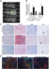
Fig. 1.
Fig. 1.
MUC1 regulates in vivo glucose uptake and expression of glycolytic genes. (A) Glucose uptake by tumors formed by orthotopically implanted S2-013.Neo.shScr (scrambled control), S2-013.MUC1.shScr (scrambled control), or S2-013.MUC1.shHIF-1α (HIF-1α knockdown). Orthotopic xenograft tumors in athymic nudes were imaged with IRDye 800CW 2DG. Arrowheads indicate the tumors. (B) Quantification of in vivo glucose uptake by tumors (normalized to tumor size). (C) Immunohistochemical staining of formalin-fixed paraffin-embedded tumor sections reveals increased expression of HIF-1α, GLUT1, and LDHA and increased nuclear Ki67 staining in S2-013.MUC1 cells in comparison with S2-013.Neo cells. Arrowheads point to nuclear Ki67 staining. (D) Immunofluorescence staining for hypoxia (EF5, red), HIF-1α (green), and nuclei (DAPI) in tumor sections from S2-013.Neo, S2-013.MUC1, and S2-013.MUC1.shHIF-1α–implanted mice (*P < 0.05 and **P < 0.01).
Fig. 2.
Fig. 2.
Interaction of MUC1.CT with HIF-1α. (A) PLA with Abs against MUC1.CT (CT2) and HIF-1α demonstrate increased interaction of MUC1 with HIF-1α under conditions of MUC1 expression and 6 h CoCl2 treatment. Arrowheads point to the interaction spots. Right: 3D localization of interaction spots in S2-013.MUC1 cells under CoCl2 treatment. (B) Quantification of interaction spots per cell in S2-013.Neo, S2-013.MUC1, FG.shScr, and FG.shMUC1 cells under conditions of 6 h CoCl2 treatment or control. (C) Coimmunoprecipitation of HIF-1α along with MUC1 from Nonidet P-40 extracts of 6 h CoCl2 treated or control S2-013.MUC1 cells. Immunoprecipitations with isotype IgG were performed as a control (*P < 0.05, **P < 0.01, and ***P < 0.001).
Fig. 3.
Fig. 3.
MUC1 facilitates HIF-1α recruitment to glycolytic gene promoters. (A) Occupancy of ENO1 and PGM2 promoters by HIF-1α was confirmed by ChIP using anti–HIF-1α Ab or IgG control, followed by qPCR analysis. Enrichment of proximal and distal ENO1 and PGM2 promoter regions from S2-013.Neo and S2-013.MUC1 cells under normoxic or hypoxic conditions (1% O2, 6 h) was compared with that from S2-013.Neo under normoxic conditions. (B) Coimmunoprecipitation of p300 along with MUC1 from Nonidet P-40 extracts of 6 h hypoxia-treated or control S2-013.MUC1 cells. M, marker lane; WCL, whole-cell lysate. (C) Occupancy of ENO1 and PGM2 promoters by p300 was confirmed by ChIP using anti-p300 Ab or IgG control, followed by qPCR analysis. Enrichment of proximal and distal ENO1 and PGM2 promoter regions from S2-013.Neo and S2-013.MUC1 cells under normoxic or hypoxic conditions was compared with that from S2-013.Neo under normoxic conditions. (D) ChIP assays were performed with IgG or anti-acetylated H3K9 (H3K9-Ac) antibody. (E) Immunoblotting to determine the total cellular levels of acetylated lysine 9 residue in histone 3 (H3K9-Ac) and the protein expression levels of histone 3 (H3) and MUC1 under hypoxic conditions in S2-013.Neo and S2-013.MUC1 cells. Tubulin was used as a loading control. (Normalized relative values are presented as vertical bars ± SEM; *P < 0.05, **P < 0.01, and ***P < 0.001).
Fig. 4.
Fig. 4.
MUC1 regulates metabolite levels in pancreatic cancer cells. (A) Three-dimensional principal component analysis scores plot comparing S2-013.Neo cells (black) with S2-013.MUC1 cells (red). (B) Heat map generated from the normalized-mean peak intensities for each metabolite identified from the triplicate set of 2D 1H-13C HSQC NMR experiments. The normalized-mean intensities are plotted on a color-scale from 0% (red) to 100% (green). Dendogram depicts hierarchal clustering of relative metabolite concentration changes between the S2-013.MUC1 and S2-013.Neo cells (*P < 0.05 and ***P < 0.001; metabolites in italics have lower levels of confidence in NMR peak assignment). (C) Metabolic pathway depicting the metabolites identified in the S2-013.MUC1 and S2-013.Neo cellular metabolome by the 2D 1H-13C HSQC NMR experiments. Three replicates of S2-013.MUC1 and S2-013.Neo cell cultures were used for metabolite identification. Dark green arrows correspond to metabolites with an increased concentration in S2-013.MUC1 cells relative to S2-013.Neo cells. Arrows colored red correspond to metabolites with a decreased concentration in S2-013.MUC1 cells relative to S2-013.Neo cells. Metabolic genes up- or down-regulated by MUC1 expression are indicated by green and red, respectively. Most glycolytic genes are not depicted for clarity. (D) Cells (5 × 10⁴) were seeded and cultured under normoxic or hypoxic conditions (1% O2) in regular or glucose/glutamine-deprived media for 3 d. Cell counts at day 3 are indicated by vertical bars ± SEM (n = 3; *P < 0.05 and ***P < 0.001 vs. counts of S2-013.Neo cells for that condition).

References

    1. Besmer DM, et al. Pancreatic ductal adenocarcinoma mice lacking mucin 1 have a profound defect in tumor growth and metastasis. Cancer Res. 2011;71:4432–4442. - PMC - PubMed
    1. Singh PK, Hollingsworth MA. Cell surface-associated mucins in signal transduction. Trends Cell Biol. 2006;16:467–476. - PubMed
    1. Singh PK, et al. Platelet-derived growth factor receptor beta-mediated phosphorylation of MUC1 enhances invasiveness in pancreatic adenocarcinoma cells. Cancer Res. 2007;67:5201–5210. - PubMed
    1. Tsutsumida H, et al. RNA interference suppression of MUC1 reduces the growth rate and metastatic phenotype of human pancreatic cancer cells. Clin Cancer Res. 2006;12:2976–2987. - PubMed
    1. Qu CF, et al. MUC1 expression in primary and metastatic pancreatic cancer cells for in vitro treatment by (213)Bi-C595 radioimmunoconjugate. Br J Cancer. 2004;91:2086–2093. - PMC - PubMed

Publication types