. 2012 Aug 21;109(34):13763-8.
doi: 10.1073/pnas.1204638109. Epub 2012 Aug 6.

Bodywide skipping of exons 45-55 in dystrophic mdx52 mice by systemic antisense delivery

Affiliations

Bodywide skipping of exons 45-55 in dystrophic mdx52 mice by systemic antisense delivery

Yoshitsugu Aoki et al. Proc Natl Acad Sci U S A. .

Abstract

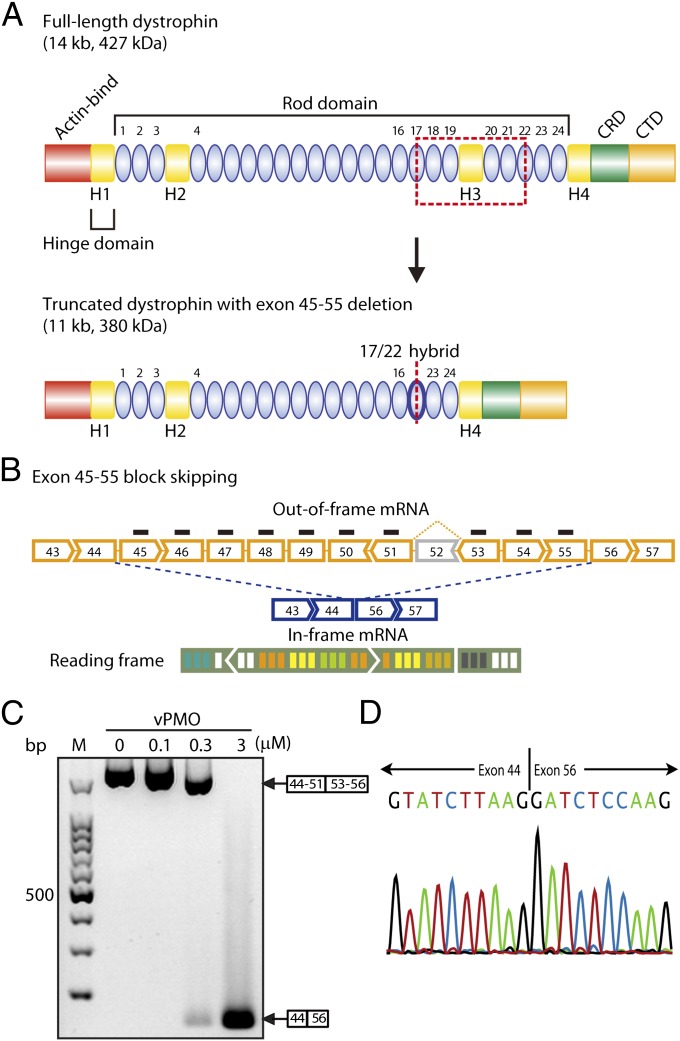
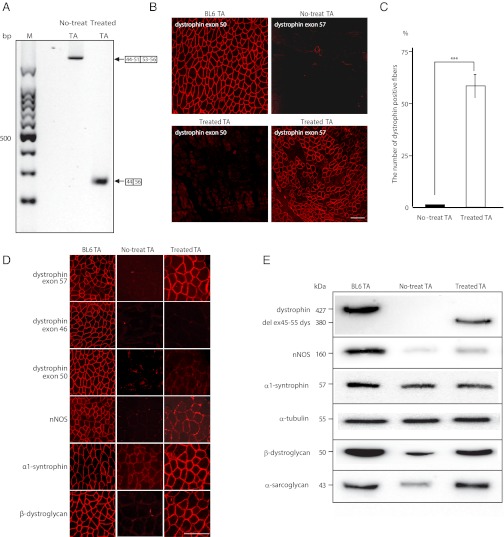
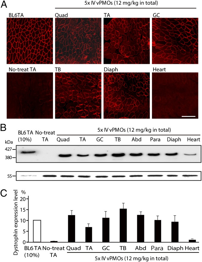
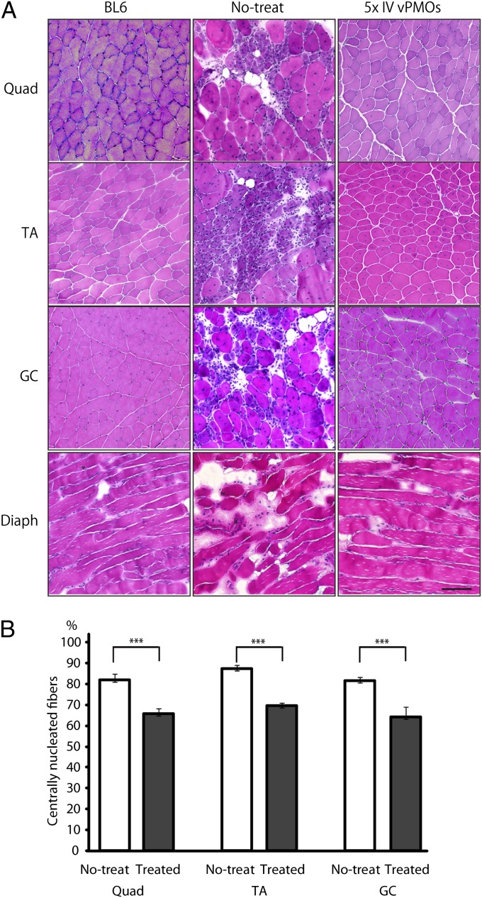
Duchenne muscular dystrophy (DMD), the commonest form of muscular dystrophy, is caused by lack of dystrophin. One of the most promising therapeutic approaches is antisense-mediated elimination of frame-disrupting mutations by exon skipping. However, this approach faces two major hurdles: limited applicability of each individual target exon and uncertain function and stability of each resulting truncated dystrophin. Skipping of exons 45-55 at the mutation hotspot of the DMD gene would address both issues. Theoretically it could rescue more than 60% of patients with deletion mutations. Moreover, spontaneous deletions of this specific region are associated with asymptomatic or exceptionally mild phenotypes. However, such multiple exon skipping of exons 45-55 has proved technically challenging. We have therefore designed antisense oligo (AO) morpholino mixtures to minimize self- or heteroduplex formation. These were tested as conjugates with cell-penetrating moieties (vivo-morpholinos). We have tested the feasibility of skipping exons 45-55 in H2K-mdx52 myotubes and in mdx52 mice, which lack exon 52. Encouragingly, with mixtures of 10 AOs, we demonstrated skipping of all 10 exons in vitro, in H2K-mdx52 myotubes and on intramuscular injection into mdx52 mice. Moreover, in mdx52 mice in vivo, systemic injections of 10 AOs induced extensive dystrophin expression at the subsarcolemma in skeletal muscles throughout the body, producing up to 15% of wild-type dystrophin protein levels, accompanied by improved muscle strength and histopathology without any detectable toxicity. This is a unique successful demonstration of effective rescue by exon 45-55 skipping in a dystrophin-deficient animal model.


Conflict of interest statement

The authors declare no conflict of interest.

Figures

Fig. 1.
Fig. 1.
Efficacy of exon 45–55 multiskipping in H2K-mdx52 cells in vitro. (A) Structure of full-length and quasidystrophin. The quasidystrophin produced by exon 45–55 deletion (skipping) has a hybrid rod repeat of rods 17 and 22. Actin-bind, actin-binding domain; CRD, cysteine-rich domain; CTD, C-terminal domain. (B) Mdx52 mouse lacks exon 52 in the mRNA of the murine Dmd gene, leading to out-of-frame products (yellow broken line). Exon 45–55 skipping with mixture vPMOs (blue broken line) restores the reading frame of dystrophin mRNA. (C) RT-PCR results after 0.1, 0.3, or 3 μM in total of mixture vPMO transfected into H2K-mdx52 myotubes as indicated. M, molecular marker; 0, no vPMO transfection. (D) Confirmation of correct exon 45–55 block skipping by direct sequencing of the PCR products. Sequencing of the most intense band shows exon 45–55 skipped dystrophin mRNA sequence.
Fig. 2.
Fig. 2.
Exon 45–55 multiskipping and rescue of dystrophin expression in mdx52 mice by local mixture-ESE2 injections. (A) Detection of exon 45–55 skipped dystrophin mRNA by RT-PCR with primers flanking exons 44 (44F1) and 56 (56R1) at 2 wk after injection of mixture-ESE2, targeting exons 45–55 except exon 52 into tibialis anterior (TA) muscles. Representative data are shown. M, molecular marker; no-treat TA, untreated TA muscles from mdx52 mice; treated TA, treated TA muscles from mdx52 mice. (B) Immunohistochemical staining of dystrophin exon 50 in the TA muscle of WT and treated mdx52 mice (Left) and dystrophin exon 57 in the TA muscle of untreated and treated mdx52 mice (Right). Representative data are shown. BL6 TA, TA muscle from a wild-type C57/BL6. (Scale bar, 100 μm.) (C) Percentage of dystrophin-positive fibers after local injections with the 10 vPMO cocktail. Data (n = 6) are presented as mean ± SD ***P < 0.001. (D) Recovery of dystrophin-associated proteins with exon 45–55 skipping. Immunohistochemical staining of dystrophin exons 57, 46, and 50, neuronal nitric oxide synthase (nNOS), α1-syntrophin, and β-dystroglycan in the TA muscle of WT, untreated, and treated mdx52 mice. Representative data are shown. BL6TA, TA muscle from a wild-type C57/BL6; no-treat TA, untreated TA muscles from mdx52 mice. (Scale bar, 100 μm.) (E) Western blotting after the mixture 10 vPMOs local injections to detect the expression of full-length dystrophin, 380-kDa quasidystrophin, nNOS, α1-syntrophin, α-tubulin, β-dystroglycan, and α-sarcoglycan in TA muscles of WT, untreated, and treated mdx52 mice. Representative data are shown.
Fig. 3.
Fig. 3.
Systemic i.v. injections of the mixture-ESE2 in mdx52 mice restore dystrophin expression in body-wide skeletal muscles. (A) Immunohistochemical staining of dystrophin exon 57 in quadriceps (Quad), TA, gastrocnemius (GC), triceps brachii (TB), diaphragm (Diaph), and heart muscles in mdx52 mice after five consecutive biweekly systemic injections of 12 mg/kg of the mixture-ESE2. Representative data are shown. BL6TA, TA muscle from wild-type C57/BL6; no-treat TA, untreated TA muscle from mdx52 mice. (Scale bar, 100 μm.) (B) Western blotting analysis with mouse monoclonal antibody DYS2 after the repeated vPMOs systemic injections into mdx52 mice. Representative data are shown. vPMO-injected muscles show 380-kDa quasidystrophin bands (Upper) and α-tubulin (Lower) in Quad, TA, GC, TB, abdominal (Abd), paraspinal (Para), Diaph, and heart muscles of treated mdx52 mice. BL6TA (10% wt/wt), TA muscle from a 10% (wt/wt) extract of wild-type C57/BL6 mice. (C) Semiquantitative analysis of dystrophin expression after AO injection. Data (n = 4) are presented as mean ± SD *P < 0.05; **P < 0.01.
Fig. 4.
Fig. 4.
Exon 45–55 skipped quasidystrophin ameliorates skeletal muscle pathology in mdx52 mice. (A) H&E staining in quadriceps (Quad), TA, gastrocnemius (GC), and diaphragm (Diaph) muscles of WT (BL6), untreated (no-treat), and treated mdx52 mice (five times i.v. vPMOs). Representative data are shown. (B) Measurement of centrally nucleated fibers (CNFs) after systemic exon 45–55 skipping (treated) and nontreated mdx52 muscles (no-treat) in Quad, TA, and GC muscles. Data (n = 4) are presented as mean ± SD ***P < 0.001.

Similar articles

Cited by

References

    1. Duchenne The pathology of paralysis with muscular degeneration (Paralysie Myosclerotique), or paralysis with apparent hypertrophy. British Medical Journal. 1867;2:541–542. - PMC - PubMed
    1. Hoffman EP, Brown RH, Jr, Kunkel LM. Dystrophin: The protein product of the Duchenne muscular dystrophy locus. Cell. 1987;51:919–928. - PubMed
    1. Koenig M, et al. The molecular basis for Duchenne versus Becker muscular dystrophy: Correlation of severity with type of deletion. Am J Hum Genet. 1989;45:498–506. - PMC - PubMed
    1. Yokota T, et al. A renaissance for antisense oligonucleotide drugs in neurology: Exon skipping breaks new ground. Arch Neurol. 2009;66:32–38. - PMC - PubMed
    1. Yokota T, et al. Efficacy of systemic morpholino exon-skipping in Duchenne dystrophy dogs. Ann Neurol. 2009;65:667–676. - PMC - PubMed

Publication types