

ERK1/2 activation is a therapeutic target in age-related macular degeneration
- PMID: 22869729
- PMCID: PMC3427082
- DOI: 10.1073/pnas.1206494109
ERK1/2 activation is a therapeutic target in age-related macular degeneration
Abstract
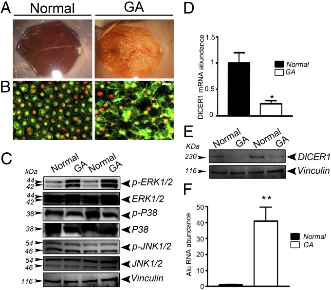
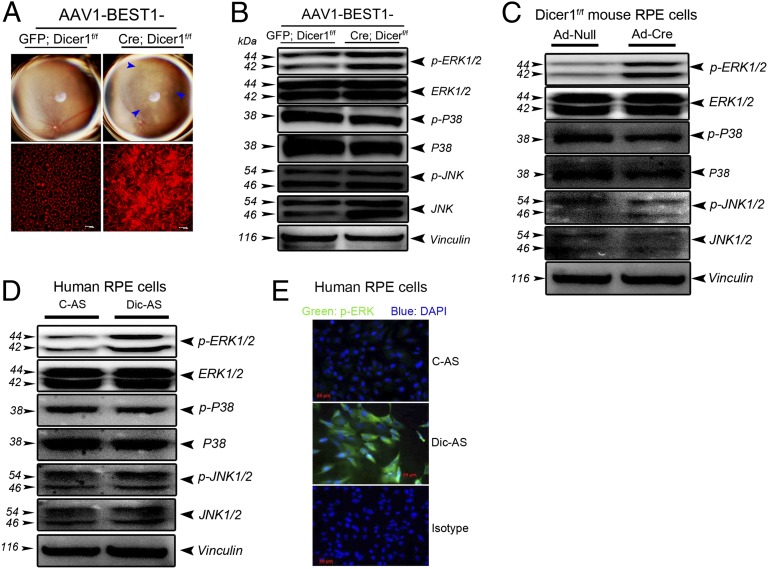
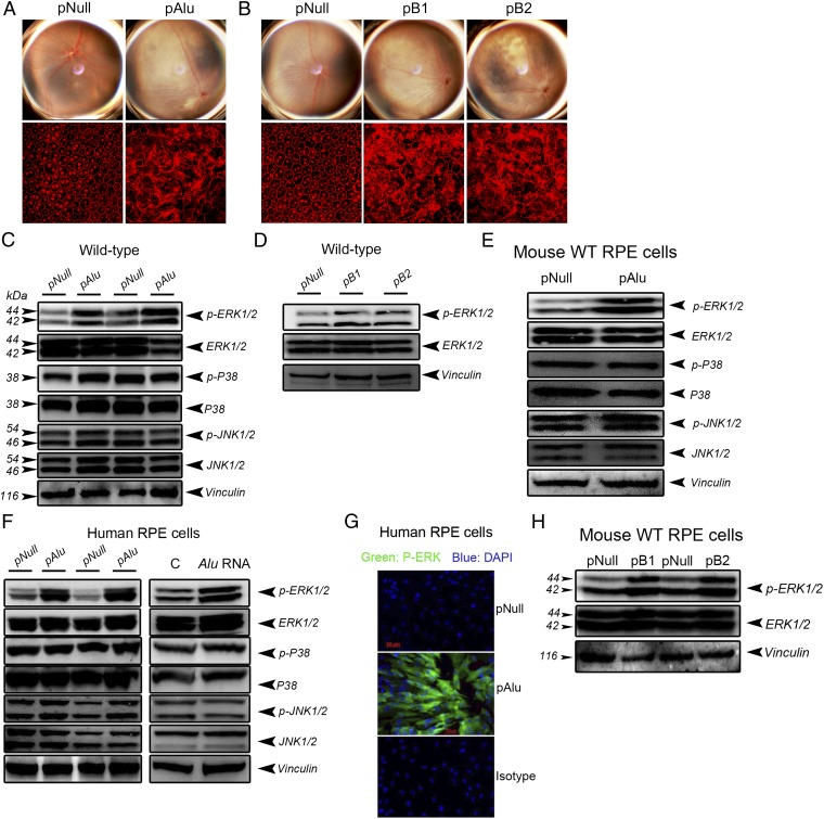
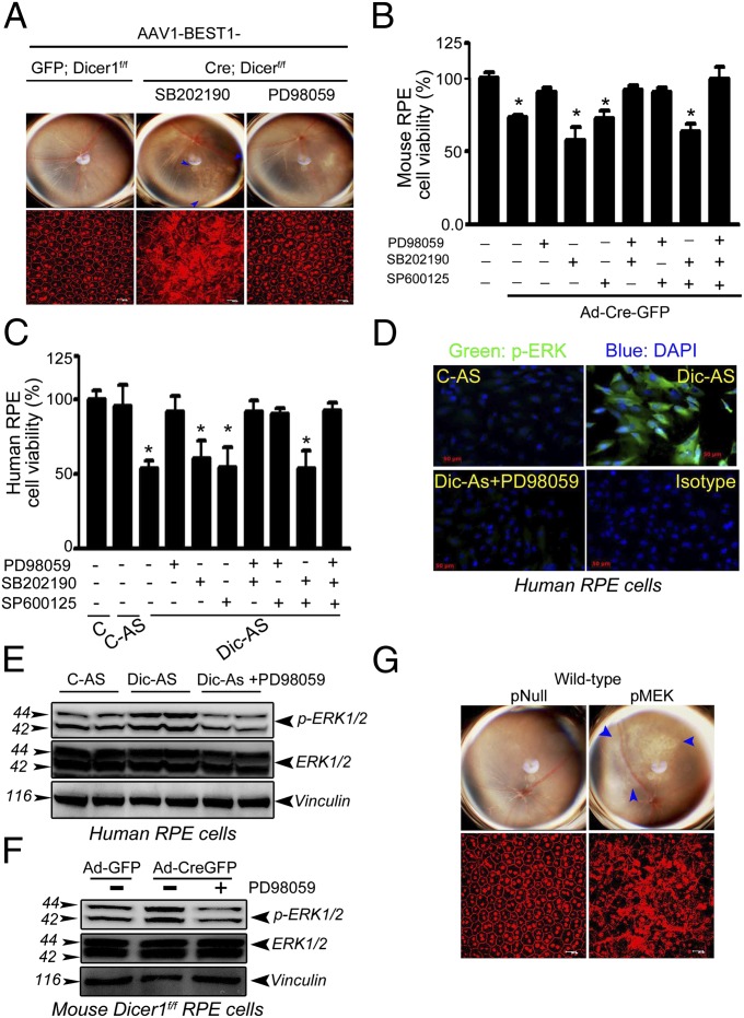
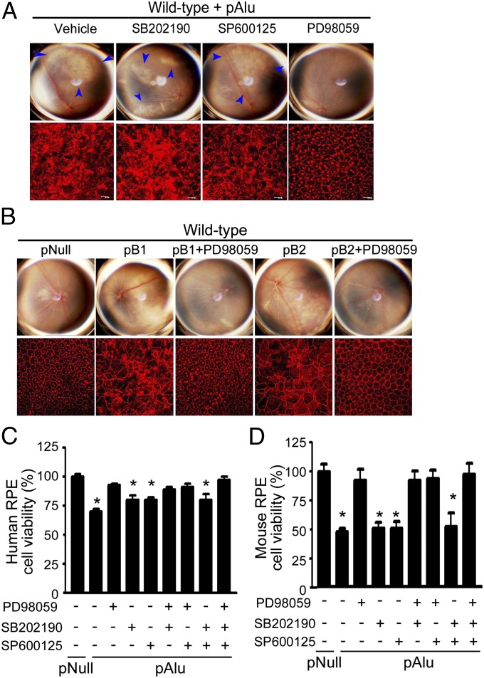
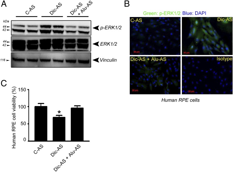
Deficient expression of the RNase III DICER1, which leads to the accumulation of cytotoxic Alu RNA, has been implicated in degeneration of the retinal pigmented epithelium (RPE) in geographic atrophy (GA), a late stage of age-related macular degeneration that causes blindness in millions of people worldwide. Here we show increased extracellular-signal-regulated kinase (ERK) 1/2 phosphorylation in the RPE of human eyes with GA and that RPE degeneration in mouse eyes and in human cell culture induced by DICER1 depletion or Alu RNA exposure is mediated via ERK1/2 signaling. Alu RNA overexpression or DICER1 knockdown increases ERK1/2 phosphorylation in the RPE in mice and in human cell culture. Alu RNA-induced RPE degeneration in mice is rescued by intravitreous administration of PD98059, an inhibitor of the ERK1/2-activating kinase MEK1, but not by inhibitors of other MAP kinases such as p38 or JNK. These findings reveal a previously unrecognized function of ERK1/2 in the pathogenesis of GA and provide a mechanistic basis for evaluation of ERK1/2 inhibition in treatment of this disease.
Conflict of interest statement
The authors declare no conflict of interest.
Figures
References
-
- Lander ES, et al. International Human Genome Sequencing Consortium Initial sequencing and analysis of the human genome. Nature. 2001;409:860–921. - PubMed
-
- Batzer MA, Deininger PL. Alu repeats and human genomic diversity. Nat Rev Genet. 2002;3:370–379. - PubMed
-
- Schmid CW, Jelinek WR. The Alu family of dispersed repetitive sequences. Science. 1982;216:1065–1070. - PubMed
-
- Dewannieux M, Esnault C, Heidmann T. LINE-mediated retrotransposition of marked Alu sequences. Nat Genet. 2003;35:41–48. - PubMed
-
- Mariner PD, et al. Human Alu RNA is a modular transacting repressor of mRNA transcription during heat shock. Mol Cell. 2008;29:499–509. - PubMed
Publication types
MeSH terms
Substances
Grants and funding
- RC1EY020442/EY/NEI NIH HHS/United States
- R01 EY017182/EY/NEI NIH HHS/United States
- RC1 EY020442/EY/NEI NIH HHS/United States
- R01 EY020672/EY/NEI NIH HHS/United States
- P30 EY003040/EY/NEI NIH HHS/United States
- T32 HL091812/HL/NHLBI NIH HHS/United States
- T32HL091812/HL/NHLBI NIH HHS/United States
- UL1 TR001998/TR/NCATS NIH HHS/United States
- R01 EY022238/EY/NEI NIH HHS/United States
- UL1 TR000117/TR/NCATS NIH HHS/United States
- R01EY001545/EY/NEI NIH HHS/United States
- R01EY020672/EY/NEI NIH HHS/United States
- R01EY017950/EY/NEI NIH HHS/United States
- UL1RR033173/RR/NCRR NIH HHS/United States
- P30EY021721/EY/NEI NIH HHS/United States
- R01GM068414/GM/NIGMS NIH HHS/United States
- TL1 RR033172/RR/NCRR NIH HHS/United States
- R01EY022238/EY/NEI NIH HHS/United States
- R01 EY018350/EY/NEI NIH HHS/United States
- R01 GM068414/GM/NIGMS NIH HHS/United States
- P30EY003040/EY/NEI NIH HHS/United States
- TL1 TR000115/TR/NCATS NIH HHS/United States
- UL1 RR033173/RR/NCRR NIH HHS/United States
- R21EY019778/EY/NEI NIH HHS/United States
- R21 EY019778/EY/NEI NIH HHS/United States
- R01EY018350/EY/NEI NIH HHS/United States
- R01 EY018836/EY/NEI NIH HHS/United States
- R01 EY001545/EY/NEI NIH HHS/United States
- K08EY021521/EY/NEI NIH HHS/United States
- K08EY021757/EY/NEI NIH HHS/United States
- R01EY017182/EY/NEI NIH HHS/United States
- K08 EY021757/EY/NEI NIH HHS/United States
- R01 EY017950/EY/NEI NIH HHS/United States
- P30 EY021721/EY/NEI NIH HHS/United States
- P30 EY014800/EY/NEI NIH HHS/United States
- K08 EY021521/EY/NEI NIH HHS/United States
- R01EY018836/EY/NEI NIH HHS/United States
LinkOut - more resources
Full Text Sources
Other Literature Sources
Medical
Molecular Biology Databases
Research Materials
Miscellaneous
