. 2012 Aug 21;109(34):13650-5.
doi: 10.1073/pnas.1203326109. Epub 2012 Aug 6.

BRCA1 and HSP90 cooperate in homologous and non-homologous DNA double-strand-break repair and G2/M checkpoint activation

Affiliations

BRCA1 and HSP90 cooperate in homologous and non-homologous DNA double-strand-break repair and G2/M checkpoint activation

Shane R Stecklein et al. Proc Natl Acad Sci U S A. .

Abstract

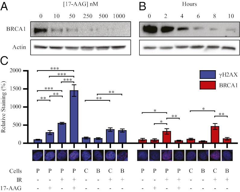
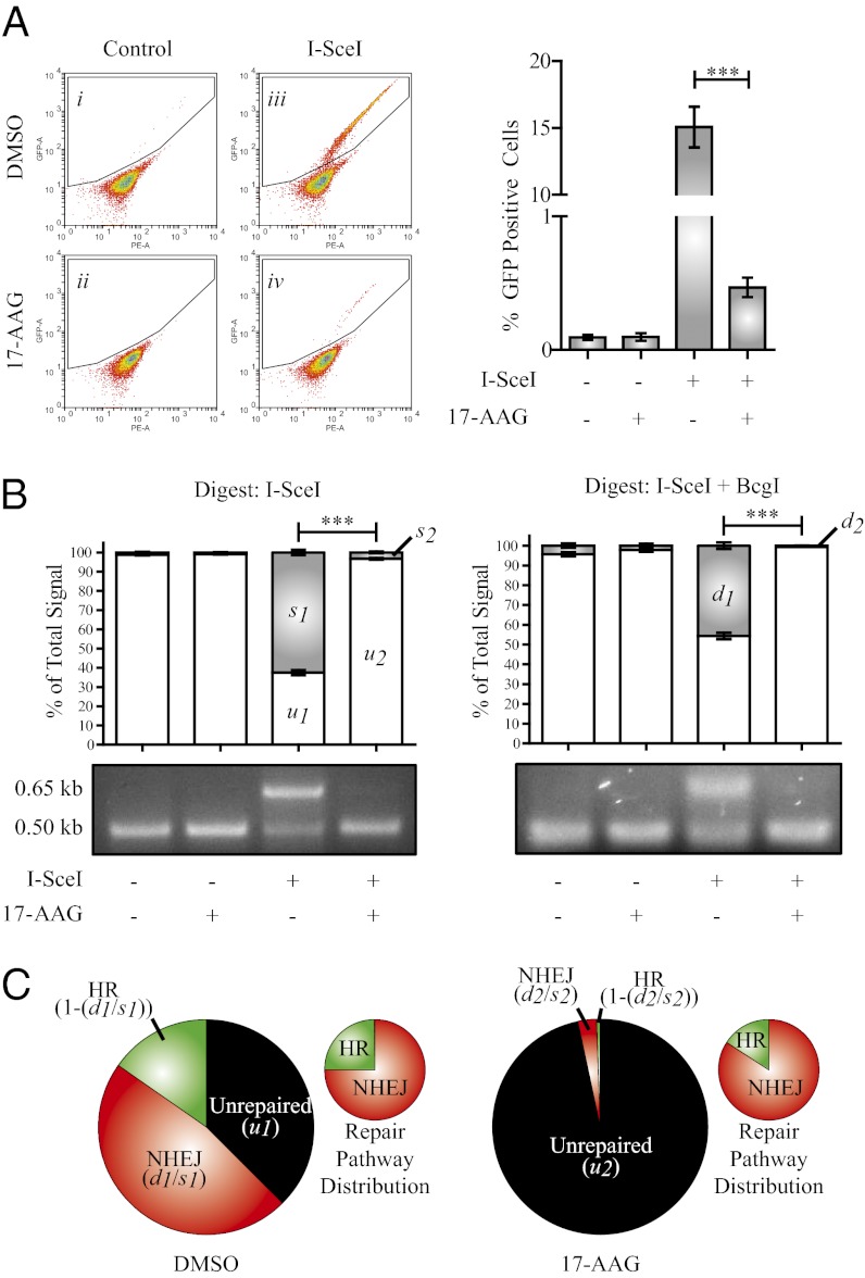
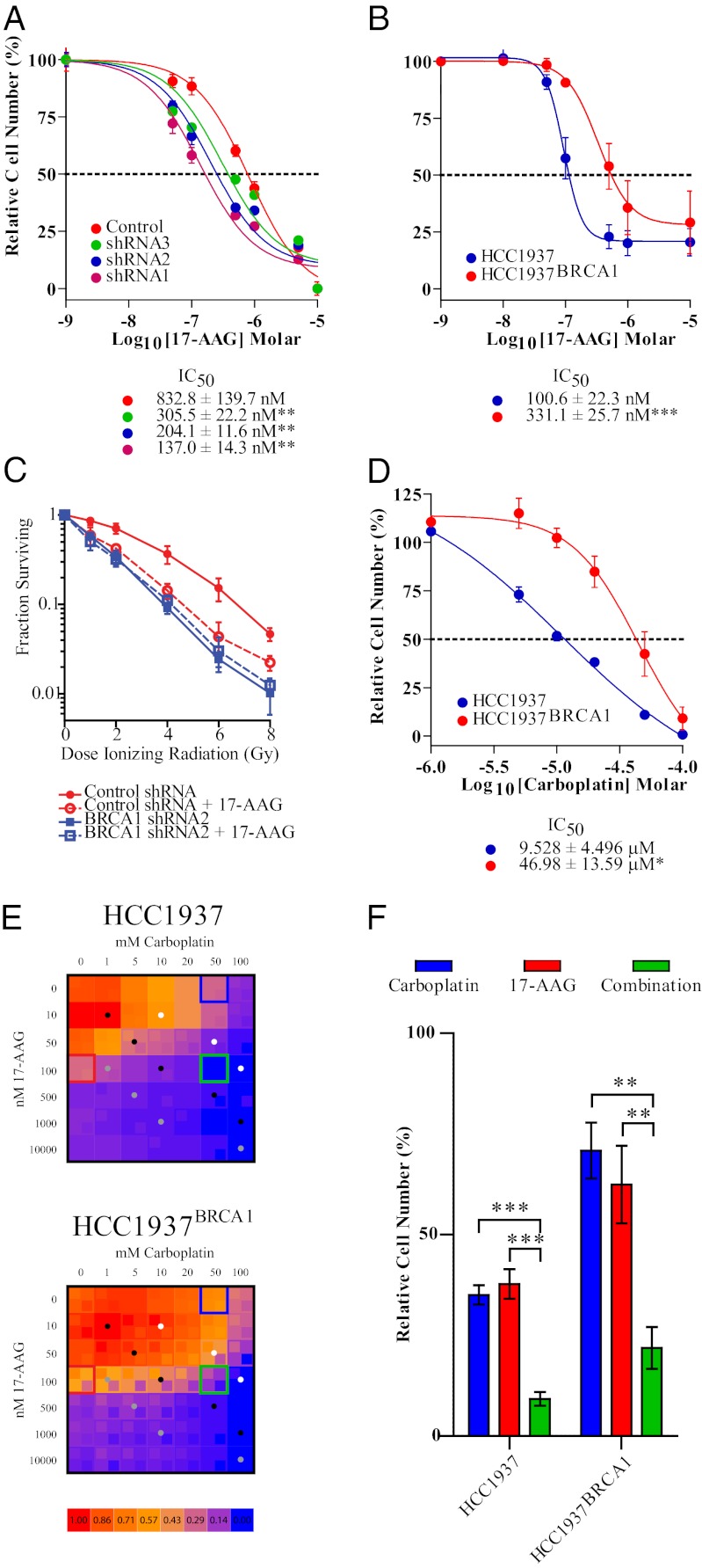
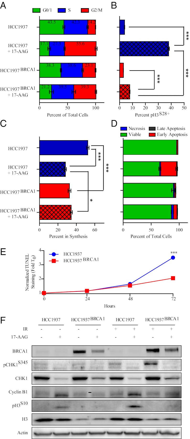
Expression of functional breast cancer susceptibility gene 1 (BRCA1) in human breast and ovarian cancers is associated with resistance to platinum-based chemotherapeutics and poly(ADP ribose) polymerase (PARP) inhibitors. BRCA1 is a nuclear tumor suppressor that is critical for resolving double-strand DNA breaks (DSBs) and interstrand crosslinks (ICLs) by homologous recombination (HR). In vitro, animal and human clinical data have demonstrated that BRCA1-deficient cancers are highly sensitive to ICL-inducing chemotherapeutic agents, are amenable to synthetic lethal approaches that exploit defects in DSB/ICL repair, and may be associated with improved survival. Conversely, high or restored expression of BRCA1 in breast and ovarian cancer is associated with therapeutic resistance and poor prognosis. There has been much interest in identifying agents that interfere with BRCA1-dependent DSB/ICL repair to restore or enhance sensitivity to cancer therapeutics. We demonstrate that the heat-shock protein 90 (HSP90) inhibitor 17-allylamino-17-demethoxygeldanamycin [17-AAG (Tanespimycin)], currently in Phase II/III clinical evaluation for several cancers, induces BRCA1 ubiquitination and proteasomal degradation, resulting in compromised repair of ionizing radiation- and platinum-induced DNA damage. We show that loss of HSP90 function abolishes BRCA1-dependent DSB repair and that BRCA1-deficient cells are hypersensitive to 17-AAG due to impaired Gap 2/Mitosis (G2/M) checkpoint activation and resultant mitotic catastrophe. In summary, we document an upstream HSP90-dependent regulatory point in the Fanconi anemia/BRCA DSB/ICL repair pathway, illuminate the role of BRCA1 in regulating damage-associated checkpoint and repair responses to HSP90 inhibitors, and identify BRCA1 as a clinically relevant target for enhancing sensitivity in refractory and/or resistant malignancies.


Conflict of interest statement

The authors declare no conflict of interest.

Figures

Fig. 1.
Fig. 1.
Inhibition of HSP90 induces degradation of BRCA1. (A) Western blots of MCF7 cells treated with indicated concentrations of 17-AAG for 8 h. (B) Western blots of MCF7 cells treated with 250 nM 17-AAG for indicated duration. (C) MCF7 cells were treated with DMSO or 250 nM 17-AAG for 24 h. Cells were exposed to 0 or 10 Gy of IR and then were fixed and immunostained for γ-H2AX or BRCA1 4 h post IR. Graphs represent the fluorescence intensity of the γ-H2AX or BRCA1 channel normalized to the DAPI channel in five random fields from one representative experiment. “Cells” designations: P, parental MCF7, C; control shRNA; B, BRCA1 shRNA1. Error bars represent SEM; *P < 0.05, **P < 0.01, and ***P < 0.001 (Student’s t test).
Fig. 2.
Fig. 2.
Inhibition of HSP90 impairs both HR and NHEJ. (A) HeLa cells stably expressing the DR–GFP reporter were electroporated with either control empty vector or a vector expressing I-SceI and were immediately plated into either DMSO or 250 nM 17-AAG for 24 h. Media was replaced after 24 h (no drug included) and cells were incubated for an additional 24 h before flow-cytometric analysis. Gated cells express GFP, indicating successful HR. Graph depicts number of GFP+ cells in three independent experiments. (B) I-SceI and I-SceI+BcgI digested PCR products. Gray bars in graph represent uncut (0.65 kb) fragment. (C) Summary of total repair capacity and repair pathway distribution in I-SceI transfected cells. Small graphs represent distribution of total repair product (HR+NHEJ). n = 3. Error bars represent SEM; *P < 0.05, **P < 0.01, and ***P < 0.001 (Student’s t test).
Fig. 3.
Fig. 3.
BRCA1 expression mediates sensitivity to 17-AAG and is associated with the ability of 17-AAG to sensitize cells to DSB-inducing agents. (A and B) MTS assay of BRCA1–shRNA-lentivirus-infected MCF7 cells (A) or HCC1937 and HCC1937BRCA1 cells (B) treated with increasing concentrations of 17-AAG. (C) Clonogenicity assay of control shRNA and shRNA2 cells pretreated with vehicle or 10 nM 17-AAG for two days and exposed to 0–6 Gy IR. Percent surviving at each dose of IR is relative to colonies formed at 0 Gy for each particular clone. (D) MTS assay of HCC1937 and HCC1937BRCA1 cells treated with increasing concentrations of carboplatin. (E) Heat maps of HCC1937 and HCC1937BRCA1 proliferation in response to various combinations of 17-AAG and carboplatin. Inlayed boxes denote mean plus SD (Upper Left) or mean minus SD (Lower Right). Colored dots denote equal ratio of carboplatin:17-AAG [10:1 (gray), 100:1 (black), 1,000:1 (white)]. (F) Proliferation of HCC1937 and HCC1937BRCA1 cells in response to 50 μM carboplatin, 100 nM 17-AAG, or both (denoted by boxes in E).
Fig. 4.
Fig. 4.
BRCA1 status regulates cell cycle progression, DNA synthesis and apoptosis in response to 17-AAG. (A) Flow-cytometric evaluation of cell cycle distribution in HCC1937 and HCC1937BRCA1 cells after 24 h of exposure to 250 nM 17-AAG. (B) pH3S28 staining in HCC1937 and HCC1937BRCA1 cells after 24 h of exposure to 250 nM 17-AAG. (C) EdU incorporation in HCC1937 and HCC1937BRCA1 cells after 24 h of exposure to 250 nM 17-AAG. (D) Annexin V staining in HCC1937 and HCC1937BRCA1 cells after 24 h of exposure to 250 nM 17-AAG. (E) TUNEL staining in HCC1937 and HCC1937BRCA1 cells after 24, 48, or 72 h of exposure to 250 nM 17-AAG. (F) Western blots for cycle checkpoint and mitosis-associated proteins in HCC1937 and HCC1937BRCA1 cells after 24 h of exposure to 250 nM 17-AAG, 10 Gy IR, or both.

Similar articles

Cited by

References

    1. Ford D, Easton DF, Bishop DT, Narod SA, Goldgar DE. Breast Cancer Linkage Consortium Risks of cancer in BRCA1-mutation carriers. Lancet. 1994;343:692–695. - PubMed
    1. King MC, Marks JH, Mandell JB. New York Breast Cancer Study Group Breast and ovarian cancer risks due to inherited mutations in BRCA1 and BRCA2. Science. 2003;302:643–646. - PubMed
    1. Thompson ME, Jensen RA, Obermiller PS, Page DL, Holt JT. Decreased expression of BRCA1 accelerates growth and is often present during sporadic breast cancer progression. Nat Genet. 1995;9:444–450. - PubMed
    1. Hall JM, et al. Linkage of early-onset familial breast cancer to chromosome 17q21. Science. 1990;250:1684–1689. - PubMed
    1. Friedman LS, et al. Confirmation of BRCA1 by analysis of germline mutations linked to breast and ovarian cancer in ten families. Nat Genet. 1994;8:399–404. - PubMed

Publication types

LinkOut - more resources