. 2012 Aug 21;109(34):13805-10.
doi: 10.1073/pnas.1206187109. Epub 2012 Aug 6.

Viroid RNA redirects host DNA ligase 1 to act as an RNA ligase

Affiliations

Viroid RNA redirects host DNA ligase 1 to act as an RNA ligase

María-Ángeles Nohales et al. Proc Natl Acad Sci U S A. .

Abstract

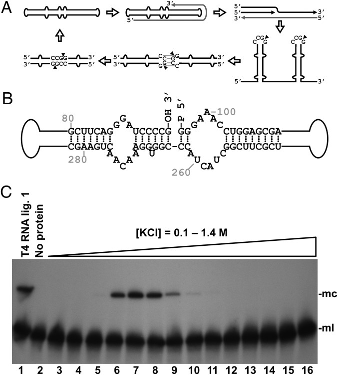
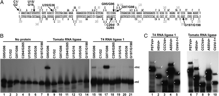
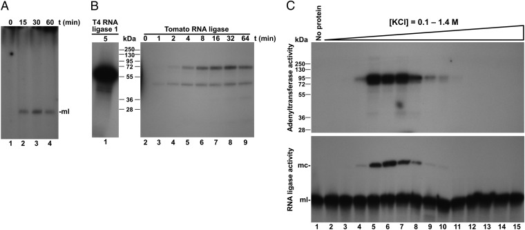
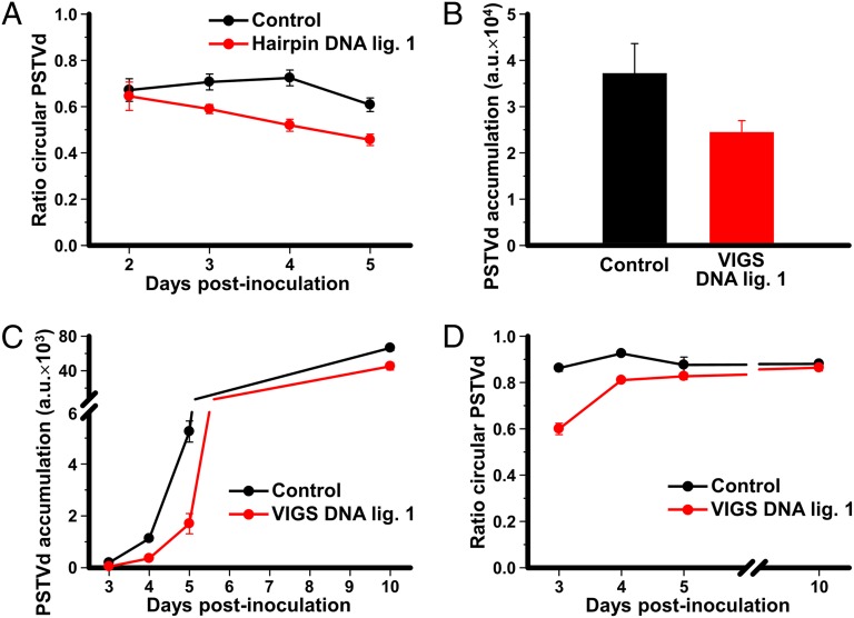
Viroids are a unique class of noncoding RNAs: composed of only a circular, single-stranded molecule of 246-401 nt, they manage to replicate, move, circumvent host defenses, and frequently induce disease in higher plants. Viroids replicate through an RNA-to-RNA rolling-circle mechanism consisting of transcription of oligomeric viroid RNA intermediates, cleavage to unit-length strands, and circularization. Though the host RNA polymerase II (redirected to accept RNA templates) mediates RNA synthesis and a type-III RNase presumably cleavage of Potato spindle tuber viroid (PSTVd) and closely related members of the family Pospiviroidae, the host enzyme catalyzing the final circularization step, has remained elusive. In this study we propose that PSTVd subverts host DNA ligase 1, converting it to an RNA ligase, for the final step. To support this hypothesis, we show that the tomato (Solanum lycopersicum L.) DNA ligase 1 specifically and efficiently catalyzes circularization of the genuine PSTVd monomeric linear replication intermediate opened at position G95-G96 and containing 5'-phosphomonoester and 3'-hydroxyl terminal groups. Moreover, we also show a decreased PSTVd accumulation and a reduced ratio of monomeric circular to total monomeric PSTVd forms in Nicotiana benthamiana Domin plants in which the endogenous DNA ligase 1 was silenced. Thus, in a remarkable example of parasitic strategy, viroids reprogram for their replication the template and substrate specificity of a DNA-dependent RNA polymerase and a DNA ligase to act as RNA-dependent RNA polymerase and RNA ligase, respectively.


Conflict of interest statement

The authors declare no conflict of interest.

Figures

Fig. 1.
Fig. 1.
Identification of a tomato RNA ligase activity circularizing the PSTVd linear replication intermediate. (A) Scheme of the asymmetric rolling-circle replication mechanism of PSTVd, including the proposed processing model of viroid oligomeric RNA (14, 15). Black and gray lines represent (+) and (−) PSTVd RNAs, respectively. Arrowheads indicate cleavage sites. (B) Structure of part of the central domain of the PSTVd monomeric linear intermediate, opened between positions G95 and G96 with 5′-phosphomonoester and 3′-hydroxyl termini, which is the physiological substrate circularized by a tomato activity. (C) Tomato RNA ligase activity circularizing the linear PSTVd replication intermediate. Monomeric linear PSTVd opened between positions G95 and G96 and containing 5′-phosphomonoester and 3′-hydroxyl termini was subjected to circularization by chromatographic protein fractions from tomato. Reaction products were separated by denaturing PAGE and PSTVd forms visualized by Northern blot hybridization. Lane 1, control with T4 RNA ligase 1; lane 2, control with no protein added; lanes 3–16, tomato chromatographic fractions eluted with a KCl gradient. The positions of the monomeric circular (mc) and monomeric linear (ml) PSTVd RNAs are shown on the right side of the image.
Fig. 2.
Fig. 2.
Substrate specificity of the viroid-circularizing activity from tomato. (A) Scheme of the minimum free-energy conformation predicted for PSTVd in which arrows indicate the positions wherein the different linear substrates are opened. (B) Monomeric linear PSTVd forms were subjected to circularization by the tomato protein fractions (lanes 8–14) or T4 RNA ligase 1 (lanes 15–21). Lanes 1–7, controls with no protein added. Reaction products were separated by denaturing PAGE, and PSTVd forms visualized by Northern blot hybridization. Positions of PSTVd monomeric circular (mc) and monomeric linear (ml) forms are indicated on the right side of the image. (C) 5′-32P–labeled monomeric linear forms of PSTVd (lanes 1 and 6), Hop stunt viroid (HSVd, lanes 2 and 7), Coconut cadang-cadang viroid (CCCVd, lanes 3 and 8), Apple scar skin viroid (ASSVd, lanes 4 and 9), and Coleus blumei viroid 1 (CbVd-1, lanes 5 and 10) were incubated with T4 RNA ligase 1 (lanes 1–5) or the most active tomato protein fraction eluted from the heparin column (lanes 6–10). Reaction products were separated by denaturing PAGE, and the gel autoradiographed. Positions of the linear and circular forms of the different viroids are indicated with asterisks and arrows, respectively.
Fig. 3.
Fig. 3.
RNA and protein adenylation during PSTVd circularization. (A and B) Monomeric linear PSTVd was incubated in the presence of [α-32P]ATP, with the most active tomato fraction eluted from the heparin column. (A) RNAs were separated by denaturing PAGE, and the gel autoradiographed. Lanes 1–4, aliquots of the reaction taken at 0, 15, 30, and 60 min. (B) Proteins were separated by SDS/PAGE, and the gel autoradiographed. Lane 1, control reaction with T4 RNA ligase 1. Lanes 2–9, aliquots of the reaction taken at 0, 1, 2, 4, 8, 16, 32, and 64 min. (C) The tomato chromatographic fractions were simultaneously assayed for protein adenylation and PSTVd circularization. In the adenylation assay (Upper), reaction products were separated by SDS/PAGE, and the gel autoradiographed. In the circularization assay (Lower), reaction products were separated by denaturing PAGE, and PSTVd detected by Northern blot hybridization. Lane 1, control with no protein added. Lanes 2–15, tomato chromatographic fractions eluted with increasing concentrations of KCl. The positions of some protein markers with their sizes in kilodaltons, and the positions of monomeric circular (mc) and monomeric linear (ml) PSTVd forms are indicated.
Fig. 4.
Fig. 4.
Effect of DNA ligase 1 silencing on the in vivo accumulation and circularization of PSTVd. (A) Interference with circularization of PSTVd RNA caused by the expression of a DNA ligase 1 hairpin. A dimeric PSTVd construct was coagroinoculated in N. benthamiana with either a tomato DNA ligase 1 hairpin construct to silence the endogenous gene (red), or with the empty vector (black). RNAs were purified from the agroinfiltrated areas of triplicate plants and separated by denaturing PAGE. Monomeric circular and linear PSTVd forms were visualized by Northern blot hybridization and quantified by phosphorimetry. The plots represent the ratio of monomeric circular to total monomeric PSTVd forms (circular plus linear) at several days postinoculation (dpi). (B–D) Interference with PSTVd accumulation and circularization caused by the expression of a VIGS construct specific for the endogenous DNA ligase 1. (B) A dimeric PSTVd construct was coagroinfiltrated in N. benthamiana 16c plants constitutively expressing GFP with either a Tobacco rattle virus construct to induce VIGS of DNA ligase 1 and GFP (red histogram) or a control construct to induce VIGS of GFP alone (black histogram). At 1 mo postinfiltration, RNA was extracted from quadruplicate samples of upper noninoculated leaves, separated by denaturing PAGE, and the monomeric circular and linear PSTVd forms detected by Northern blot hybridization and quantified as indicated above. Histograms represent PSTVd accumulation in arbitrary units (a.u.). (C and D) N. benthamiana 16c plants constitutively expressing GFP were preinoculated with the VIGS vectors to induce silencing of either the endogenous DNA ligase 1 and GFP (red) or GFP alone (control, black). At 3–4 wk later, GFP silenced plants were agroinfiltrated with a construct to express a dimeric PSTVd RNA. RNA was extracted at different time intervals from the agroinoculated areas of triplicate plants, and PSTVd forms were visualized by Northern blot hybridization and quantified. (C) Plot of total PSTVd RNA accumulation (in a.u.) vs. dpi. (D) Plot of the ratio of monomeric circular to total PSTVd monomeric forms vs. dpi. All plots include bars indicating the SD of the replicate measures.

Similar articles

Cited by

References

    1. Diener TO, Raymer WB. Potato spindle tuber virus: A plant virus with properties of a free nucleic acid. Science. 1967;158:378–381. - PubMed
    1. Gross HJ, et al. Nucleotide sequence and secondary structure of potato spindle tuber viroid. Nature. 1978;273:203–208. - PubMed
    1. Semancik JS, Weathers LG. Exocortis virus of citrus: Association of infectivity with nucleic acid preparations. Virology. 1968;36:326–328. - PubMed
    1. Flores R, Hernández C, Martínez de Alba AE, Daròs JA, Di Serio F. Viroids and viroid-host interactions. Annu Rev Phytopathol. 2005;43:117–139. - PubMed
    1. Ding B. The biology of viroid-host interactions. Annu Rev Phytopathol. 2009;47:105–131. - PubMed

Publication types