. 2012 Aug 21;109(34):13793-8.
doi: 10.1073/pnas.1205526109. Epub 2012 Aug 6.

Direct reprogramming of fibroblasts into endothelial cells capable of angiogenesis and reendothelialization in tissue-engineered vessels

Affiliations

Direct reprogramming of fibroblasts into endothelial cells capable of angiogenesis and reendothelialization in tissue-engineered vessels

Andriana Margariti et al. Proc Natl Acad Sci U S A. .

Abstract

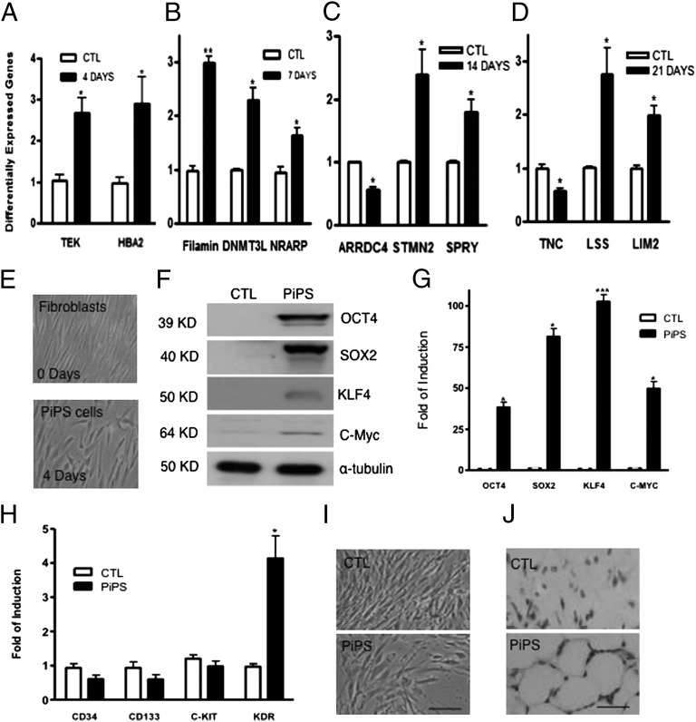
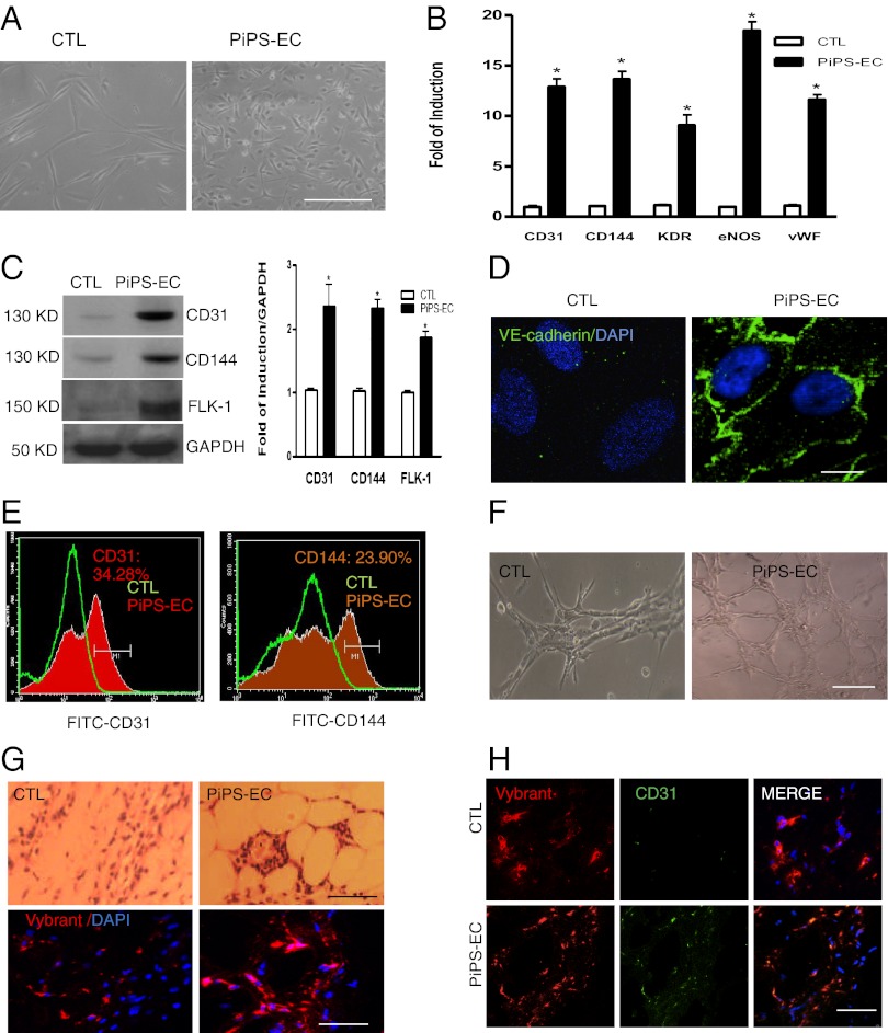
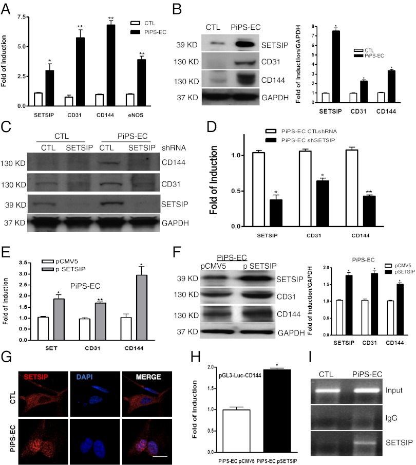
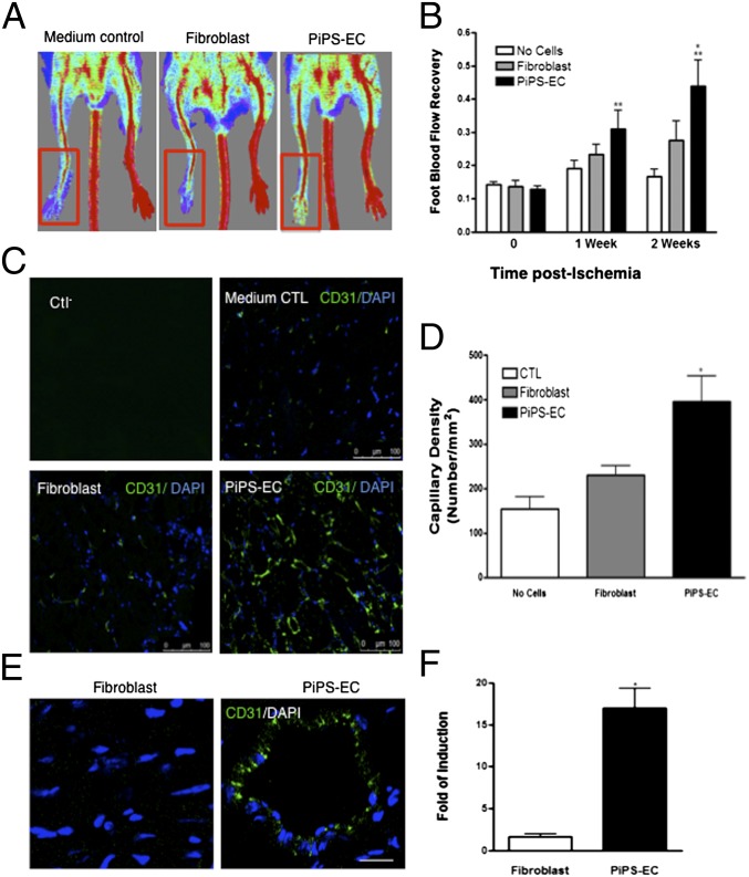
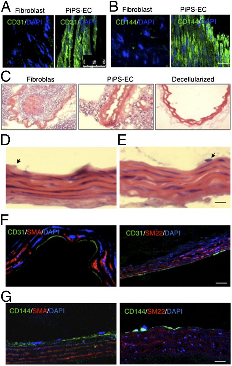
The generation of induced pluripotent stem (iPS) cells is an important tool for regenerative medicine. However, the main restriction is the risk of tumor development. In this study we found that during the early stages of somatic cell reprogramming toward a pluripotent state, specific gene expression patterns are altered. Therefore, we developed a method to generate partial-iPS (PiPS) cells by transferring four reprogramming factors (OCT4, SOX2, KLF4, and c-MYC) to human fibroblasts for 4 d. PiPS cells did not form tumors in vivo and clearly displayed the potential to differentiate into endothelial cells (ECs) in response to defined media and culture conditions. To clarify the mechanism of PiPS cell differentiation into ECs, SET translocation (myeloid leukemia-associated) (SET) similar protein (SETSIP) was indentified to be induced during somatic cell reprogramming. Importantly, when PiPS cells were treated with VEGF, SETSIP was translocated to the cell nucleus, directly bound to the VE-cadherin promoter, increasing vascular endothelial-cadherin (VE-cadherin) expression levels and EC differentiation. Functionally, PiPS-ECs improved neovascularization and blood flow recovery in a hindlimb ischemic model. Furthermore, PiPS-ECs displayed good attachment, stabilization, patency, and typical vascular structure when seeded on decellularized vessel scaffolds. These findings indicate that reprogramming of fibroblasts into ECs via SETSIP and VEGF has a potential clinical application.


Conflict of interest statement

The authors declare no conflict of interest.

Figures

Fig. 1.
Fig. 1.
Different expression of genes during reprogramming and characterization of 4-day PiPS cells. Differential expression profile of genes altered according to microarray analysis during reprogramming was confirmed by real-time PCR assays on day 4 (A), day 7 (B), day 14 (C), and day 21 (D) [data are means ± SEM (n = 3); *P < 0.05, **P < 0.01]. Human fibroblasts were nucleofected with a linearized pCAG2LMKOSimO plasmid encoding the four reprogramming genes (OCT4, SOX2, KLF4, and C-MYC) or an empty vector. Images show the morphology of fibroblasts, 4-day PiPS cells (E). PiPS cells expressed the four reprogramming factors at protein (F) and mRNA levels (G) [data are means ± SEM (n = 3); *P < 0.05, ***P < 0.001]. (H) Real-time PCR assays for progenitor markers CD34, CD133, c-Kit, and KDR (VEGFR2) [data are means ± SEM (n = 3); *P < 0.05]. (I) PiPS cells were negative for alkaline phosphatase, whereas they formed capillary-like structures in in vivo Matrigel plug assays, when injected to SCID mice for 2 wk (J). (Scale bar, 50 μm.)
Fig. 2.
Fig. 2.
PiPS cells differentiate into ECs. PiPS or control cells were seeded on collagen IV-coated plates and cultured with endothelial cell growth medium-2 (EGM-2) for 6 d. (A) Images show an endothelial-like morphology for PiPS-ECs in comparison with control cells, and expressed endothelial-specific cell markers, such as CD31, CD144, KDR, eNOS, and vWF, at the mRNA [data are means ± SEM (n = 3); *P < 0.05] (B) and protein levels and quantification (C). (D) Immunoflurorescence staining showed a typical endothelial staining for CD144, and DAPI was used and stained the cell nucleus. (Scale bar, 25 μm.) (E) FACS analysis confirmed expression of CD31 and CD144. Representative images show vascular-like tubes in vitro (F) and in vivo (G, Upper) in Matrigel plug assays. PiPS-ECs labeled with Vybrant (red) before the s.c. injection in SCID mice (G, Lower) confirmed the presence of labeled human cells 2 wk later. The PiPS-ECs stained positive for Vybrant and CD31, whereas the control cells were positive only for the Vybrant but not for CD31 (H). (Scale bar, 50 μm.)
Fig. 3.
Fig. 3.
SETSIP is involved in differentiation of PiPS-ECs. Real-time PCR shows SETSIP parallel expression with EC markers at the mRNA [A; data are means ± SEM (n = 3); *P < 0.05, **P < 0.01] and protein (B) levels and quantification (Right), in PiPS-ECs at day 6 of differentiation. SETSIP was knocked down by shRNA in CTL and PiPS-ECs at day 3 of differentiation, showing a suppression in endothelial markers assessed on day 6 at protein (C) and mRNA levels [D; data are means ± SEM (n = 3); *P < 0.05, **P < 0.01]. SETSIP overexpression using pCMV5-SETSIP construct (p SETSIP) or empty vector pCMV5 in PiPS-ECs during EC differentiation at day 4, showing further induction in EC marker expression at mRNA [E; data are means ± SEM (n = 3); *P < 0.05, **P < 0.01] and protein levels, and quantification (F), when assessed at day 6. Immunostaining shows translocation of SETSIP to the cell nucleus during PiPS-ECs differentiation at day 6; DAPI was used and stained the cell nucleus (G). (Scale bar, 50 μm.) Luciferase assays were performed at day 4 during PiPS-ECs differentiation, showing an increased promoter activity for VE-cadherin in the presence of the SETSIP at day 6 of differentiation (H; data are means ± SEM (n = 3); *P < 0.05]. ChIP assays revealed that SETSIP binds directly to the VE-cadherin gene promoter at region (−864 to −1152) nt upstream of the transcription initiation site in 6-day differentiated PiPS-ECs (I).
Fig. 4.
Fig. 4.
PiPS-ECs improved neovascularization and blood flow recovery in a hindlimb ischemic model. PiPS-ECs, fibroblasts, or medium control (no cells) were injected i.m. into adductors of an ischemic model of SCID mice. (A) Representative color laser Doppler images of superficial blood flow (BF) in lower limbs taken 2 wk after ischemia induction. (B) Line graph shows the time course of postischemic foot BF recovery (calculated as the ratio between ischemic foot BF and contralateral foot BF) in mice given medium as control, fibroblasts, and PiPS-ECs. Statistical analysis showed significantly higher foot BF recovery for PiPS-ECs in comparison with both “no cells” control and fibroblasts at weeks 1 and 2 [data are means ± SEM (n = 6)]. Week 1: **P < 0.01, PiPS-EC vs. “no cells” control; **P < 0.01, PiPS-EC vs. fibroblasts. Week 2: **P < 0.01, PiPS-EC vs “no cell” control; *P < 0.05 PiPS-EC vs. fibroblasts. No significant differences were detected when fibroblasts were compared with “no cells” control. (C) Sections of adductors muscles were stained with CD31 antibody, and capillary density was expressed as the capillary number per mm2 [D; data are means ± SEM (n = 3); *P < 0.05]. (Scale bar, 100 μm.) (E) PiPS-ECs displayed an enhanced engraftment ability compared with fibroblasts when stained and quantified with a human-specific CD31 antibody at six randomly selected microscopic fields (at ×100) [F; data are means ± SEM (n = 3); *P < 0.05]. (Scale bar, 50 μm.)
Fig. 5.
Fig. 5.
PiPS-ECs displayed endothelial properties ex vivo. PiPS-ECs or fibroblasts were seeded on decellularized vessel scaffolds in a specially constructed bioreactor and harvested on day 5. (A and B) En face positive staining for endothelial markers CD31 and CD144 is shown in the PiPS-EC. (Scale bar, 50 μm.) (C) H&E staining showed that fibroblast-seeded vessels were almost occluded, whereas PiPS-ECs-seeded scaffolds presented with a normal vessel morphology. (D and E) Cross-sections with H&E staining of the double-seeded scaffolds with selected PiPS cells, which were induced to be differentiated into smooth-muscle cells and ECs, showing a native-like vessel architecture with multiple layers of smooth-muscle cells and a monolayer of ECs. Arrows indicate an endothelial-like cell. (F and G) Double-seeded PiPS cell-derived vessels were fixed and stained positive for EC markers, such as CD31 and CD144, and for smooth-muscle markers, such as SMA and SM22. (Scale bar, 50 μm.)

Comment in

References

    1. Takahashi K, Okita K, Nakagawa M, Yamanaka S. Induction of pluripotent stem cells from fibroblast cultures. Nat Protoc. 2007;2:3081–3089. - PubMed
    1. Takahashi K, Yamanaka S. Induction of pluripotent stem cells from mouse embryonic and adult fibroblast cultures by defined factors. Cell. 2006;126:663–676. - PubMed
    1. Okita K, Nakagawa M, Hyenjong H, Ichisaka T, Yamanaka S. Generation of mouse induced pluripotent stem cells without viral vectors. Science. 2008;322:949–953. - PubMed
    1. Yu J, et al. Induced pluripotent stem cell lines derived from human somatic cells. Science. 2007;318:1917–1920. - PubMed
    1. Maherali N, et al. Directly reprogrammed fibroblasts show global epigenetic remodeling and widespread tissue contribution. Cell Stem Cell. 2007;1:55–70. - PubMed

Publication types

MeSH terms