. 2012 Sep 15;189(6):2824-32.
doi: 10.4049/jimmunol.1201114. Epub 2012 Aug 6.

The chemokine receptors CXCR1 and CXCR2 couple to distinct G protein-coupled receptor kinases to mediate and regulate leukocyte functions

Affiliations

The chemokine receptors CXCR1 and CXCR2 couple to distinct G protein-coupled receptor kinases to mediate and regulate leukocyte functions

Sandeep K Raghuwanshi et al. J Immunol. .

Abstract

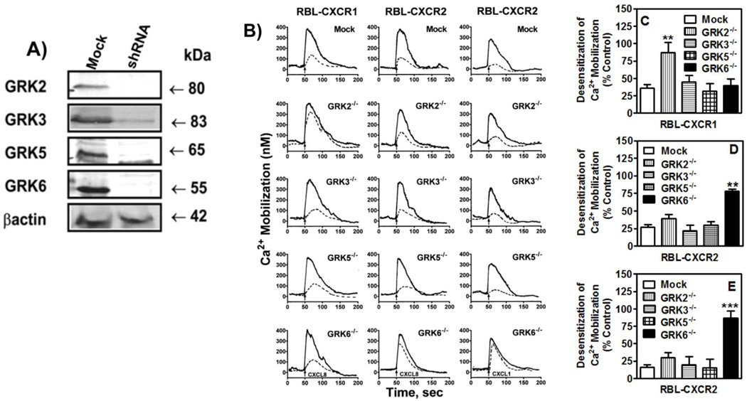
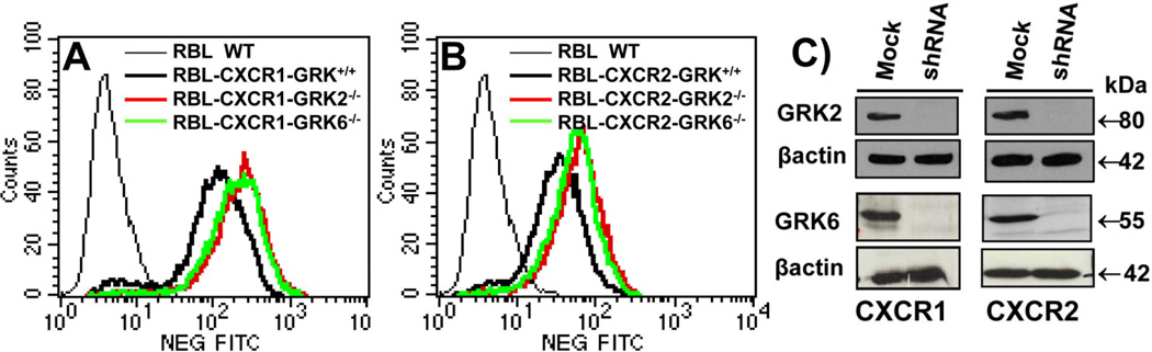
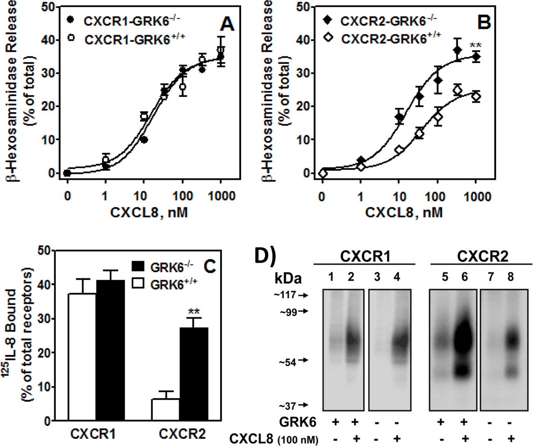
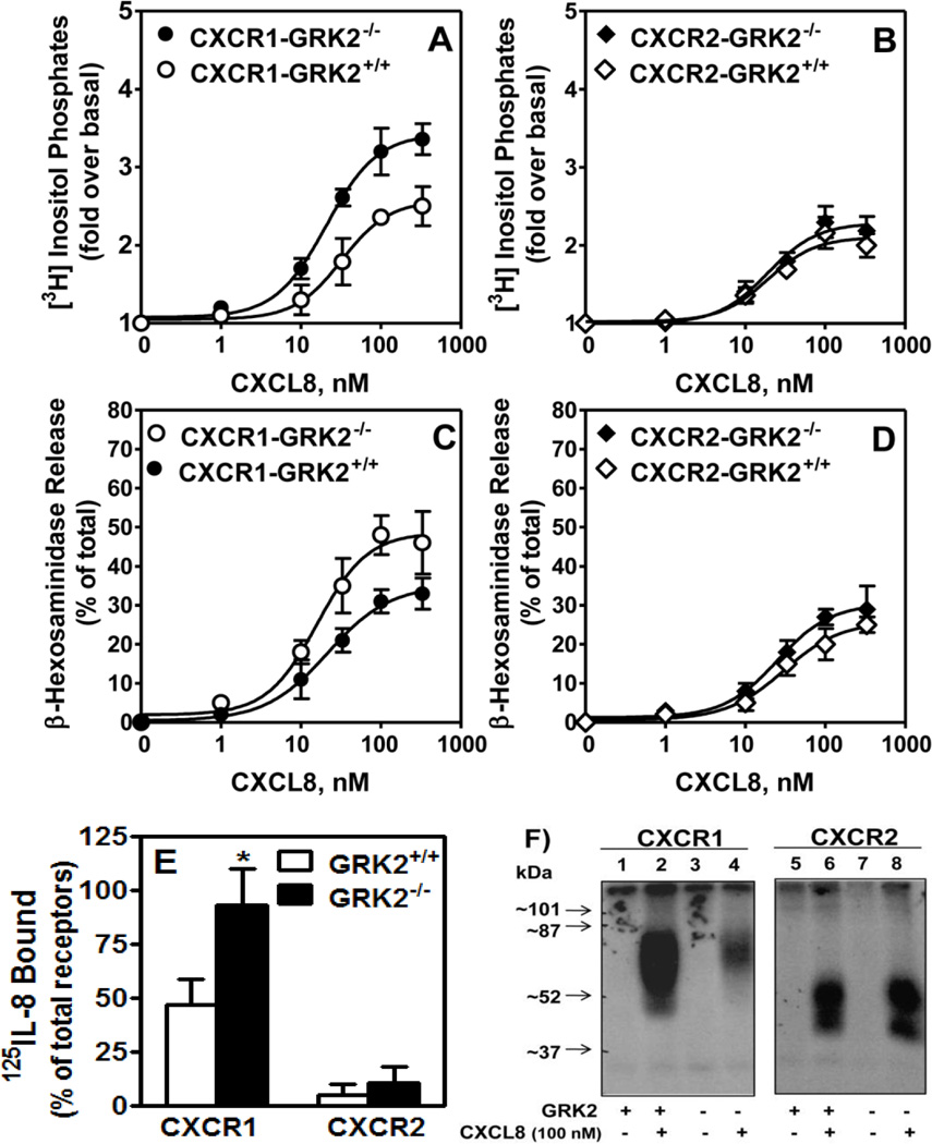
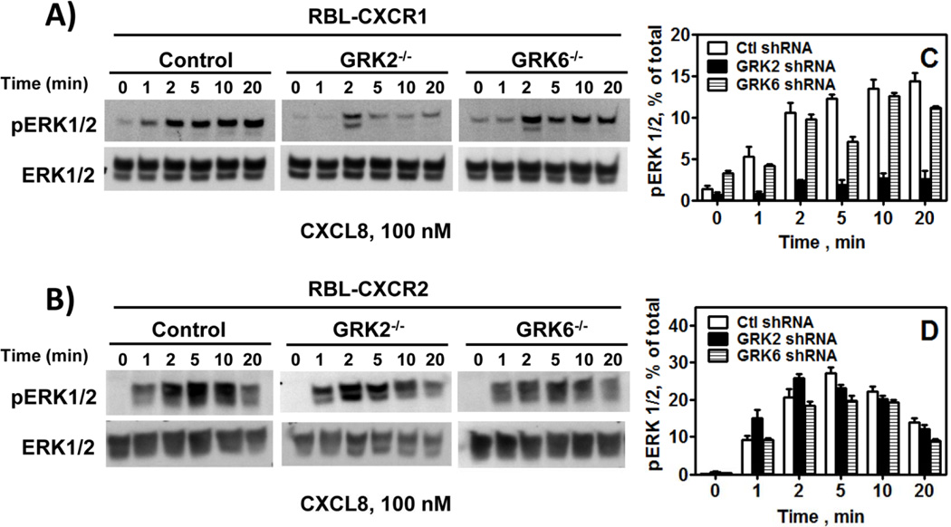
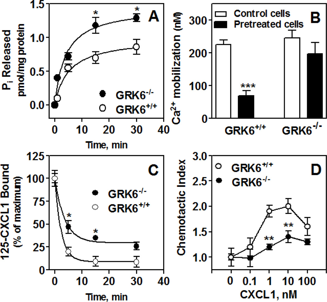
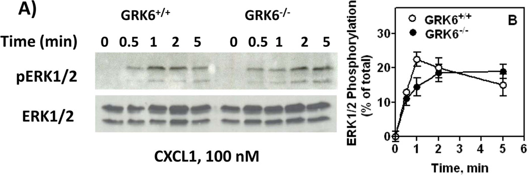
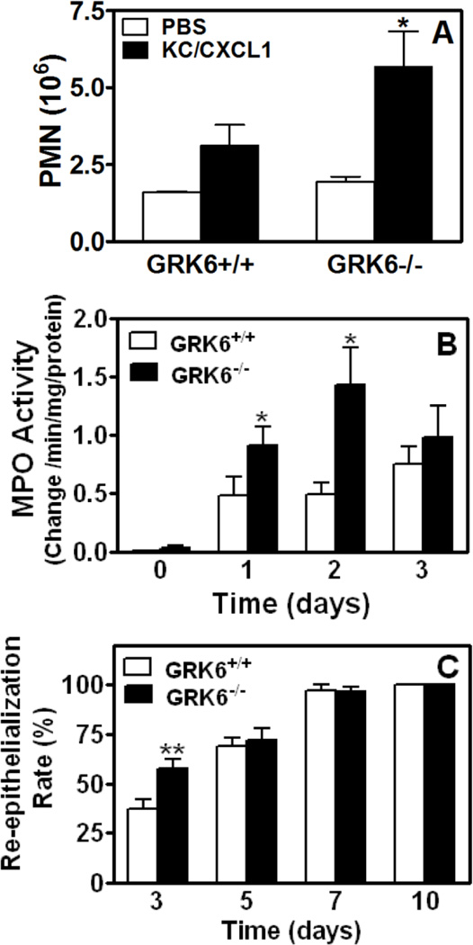
The chemokine receptors, CXCR1 and CXCR2, couple to Gαi to induce leukocyte recruitment and activation at sites of inflammation. Upon activation by CXCL8, these receptors become phosphorylated, desensitized, and internalized. In this study, we investigated the role of different G protein-coupled receptor kinases (GRKs) in CXCR1- and CXCR2-mediated cellular functions. To that end, short hairpin RNA was used to inhibit GRK2, 3, 5, and 6 in RBL-2H3 cells stably expressing CXCR1 or CXCR2, and CXCL8-mediated receptor activation and regulation were assessed. Inhibition of GRK2 and GRK6 increased CXCR1 and CXCR2 resistance to phosphorylation, desensitization, and internalization, respectively, and enhanced CXCL8-induced phosphoinositide hydrolysis and exocytosis in vitro. GRK2 depletion diminished CXCR1-induced ERK1/2 phosphorylation but had no effect on CXCR2-induced ERK1/2 phosphorylation. GRK6 depletion had no significant effect on CXCR1 function. However, peritoneal neutrophils from mice deficient in GRK6 (GRK6(-/-)) displayed an increase in CXCR2-mediated G protein activation but in vitro exhibited a decrease in chemotaxis, receptor desensitization, and internalization relative to wild-type (GRK6(+/+)) cells. In contrast, neutrophil recruitment in vivo in GRK6(-/-) mice was increased in response to delivery of CXCL1 through the air pouch model. In a wound-closure assay, GRK6(-/-) mice showed enhanced myeloperoxidase activity, suggesting enhanced neutrophil recruitment, and faster wound closure compared with GRK6(+/+) animals. Taken together, the results indicate that CXCR1 and CXCR2 couple to distinct GRK isoforms to mediate and regulate inflammatory responses. CXCR1 predominantly couples to GRK2, whereas CXCR2 interacts with GRK6 to negatively regulate receptor sensitization and trafficking, thus affecting cell signaling and angiogenesis.


Figures

Figure 1
Figure 1. Transient inhibition of GRK2, GRK3, GRK5 and GRK6 expression in RBL-2H3 cells and their effects in CXCR1 and CXCR2 desensitization
A) RBL cells (5 × 10⁵ cells/well) were transfected with 20 µg of control (mock) or siRNA specific for GRK2, GRK3, GRK5 or GRK6. Forty-eight h post transfection, cells were lysed and analyzed by immunoblotting. B) For receptor desensitization, GRK deficient and control (mock transfected) RBL cells (5×10⁶ cells) expressing CXCR1 or CXCR2 were loaded with Indo-1 in the presence or absence of 10 nM CXCL8 (left and middle columns) or CXCL1 (right column) for 30 min and assayed for CXCL8 or CXCL1 (10 nM) induced intracellular Ca2+ mobilization. Traces shown are representative of 3–5 experiments. C, D and E) Desensitization were determined as percentage of control which is the peak of intracellular Ca2+ mobilization obtained in the absence of pretreatment. Data shown are average of at least 3 traces. Statistical difference is evaluated by Student’s t test. **P<0.01; *** P < 0.001.
Figure 2
Figure 2. Stable knockdown of GRK2 and GRK6 expression in RBL-2H3 cells stably expressing CXCR1 and CXCR2
A & B) A representative histogram of FACS analysis showing surface expression of CXCR1 (A) and CXCR2 (B) in RBL-2H3 cells after staining with CXCR1 or CXCR2 specific antibodies. B) RBL-2H3 cells expressing CXCR1 (left panel) or CXCR2 (right panel) were transfected with scrambled shRNA control lentivirus (mock) or shRNA lentivirus specific for GRK2 or GRK6 knockdown. Puromycin resistant cells were selected and single clones were generated and analyzed by immunoblotting.
Figure 3
Figure 3. GRK6 inhibition enhances CXCR2-mediated exocytosis and decreases receptor phosphorylation and internalization
A & B) For β-hexosaminidase release, cells (50,000/well) were cultured overnight, washed with HEPES-buffered saline and stimulated with different concentrations of CXCL8 for 10 min. Supernatant (15 µl) was removed, and β-hexosaminidase release was measured. Data are represented as percentage of total β-hexosaminidase release from cell lysates. The experiments were repeated four times with similar results. **P<0.01. C) For receptor internalization, cells (0.5×10⁶/well) were treated with CXCL8 (100 nM) or vehicle control for 60 min and assayed for 125I-CXCL8 binding. Data are represented as percentage of total 125I-CXCL8 bound to control (untreated) cells. **P<0.01. D) For receptor phosphorylation, 32P-labeled GRK6+/+ (lanes 1 & 2) and GRK6−/− (lanes 3 & 4) RBL cells (5×10⁶cells/60 mm plate) expressing CXCR1 (left panels) or CXCR2 (right panels) were incubated for 5 min with (lanes 2 & 4) or without (lanes 1 & 3) 100 nM CXCL8. Cells were lysed, immunoprecipitated with CXCR1 and CXCR2 antibodies, analyzed by SDS-PAGE, and autoradiographed. The results shown are from a representative experiment that was repeated twice.
Figure 4
Figure 4. GRK2 knockdown increases CXCR1-mediated exocytosis but decreases receptor phosphorylation and internalization
For the generation of inositol phosphates (A and B), cells (50,000/well) were cultured overnight in the presence of [3H]inositol (1 µC/ml), washed with HEPES-buffered saline, pre-incubated for 10 min at 37°C with HEPES-buffered saline containing 10 mM LiCl in a total volume of 200µl and stimulated with different concentrations of CXCL8 for 10 min. The supernatant was used to determine the release of inositol phosphates. Data are represented as the fold stimulation over basal. β-hexosaminidase release (C & D) was measured as described in the legend of Fig. 3A and B. Data are represented as fold over basal PI (A & B), or percentage of total β-hexosaminidase release from cell lysates (C & D). Data shown are average of three experiments performed in triplicate. Receptor internalization (E) and phosphorylation (F) were measured as described in the legend of Figure 3. The results are average (E) or a representative (F) of three experiments. * p<0.05, Student’s t test.
Figure 5
Figure 5
Effect of GRK2 and GRK6 knockdown in CXCR1 and CXCR2-induced ERK1/2 phosphorylation: RBL-2H3 cells were stimulated with CXCL8 (100 nM) for 0–20 min. ERK1/2 phosphorylation and total ERK were determined by Western blotting using anti-phospho-ERK1/2 (pERK1/2) and anti-total ERK1/2 (ERK1/2) antibodies, respectively. Results shown are % of total ERK and are average of 3 experiments.
Figure 6
Figure 6
CXCL1-induced GTPase activity (A), intracellular Ca2+ mobilization (B), chemotaxis (D) and receptor internalization (C) in neutrophils from mice deficient in GRK6 (GRK6−/−) relative to wild type (GRK6+/+). Zymosan-elicited peritoneal neutrophils were collected from mice deficient in GRK6 (GRK6−/−) and control littermates (GRK6+/+). A) Membranes were prepared from zymosan-elicited peritoneal neutrophils and assayed for time-dependent CXCL1-stimulated Pi released. Results shown are representative of one of two experiments performed in triplicate. *P<0.05; **P<0.01, Student’s t test. B) Cells (3 × 10⁶ cells) were Indo-1–loaded pretreated with or without CXCL1 (100 nM) and stimulated with 10 nM CXCL1. The data shown are representative of at least 3 traces. C) Cells (0.5×10⁶ cells) were treated with CXCL1 (100 nM) for different period of time and assayed for 125I-CXCL1 binding. Data are represented as percentage of total 125I-CXCL8 bound to control (untreated) cells. **P<0.01, Student’s t test. D) Cells were incubated with calcein AM ionophore for 30 min and resuspended (1.0 × 10⁵/20ml) in RPMI without phenol red. Different concentrations of CXCL8 were loaded in a neuroprobe 96-well plate. The cells were added to the top of the filter and incubated for 2 h at 37 °C. After incubation, the top of the filter was washed five times with medium and fluorescence intensity of the bottom well-plate was measured in a Perkin Elmer fluorescence microplate reader. The experiment was repeated 3 times in triplicate. **P<0.01; *** P < 0.001, Student’s t test.
Figure 7
Figure 7
CXCL1-induced ERK activity. Zymosan-elicited peritoneal neutrophils from GRK6−/− and GRK6+/+ mice were treated with CXCL1 for different periods of time. Cell lysates were assayed for ERK phosphorylation using phospho-ERK antibody. Results shown are % of total ERK and are average of 3 experiments.
Figure 8
Figure 8
Effect of GRK6 knockdown in leukocyte migration, myeloperoxidase (MPO) activity and wound re-epithelialization. A) Six-day air pouches were raised in the dorsum of 6–8 week old GRK6−/− mice and their littermates (GRK6+/+). Mice were injected with 0.5 ml PBS or PBS containing murine CXCL1 (100 pmol). Exudates were collected after 4 h and the total number of leukocytes (~90% neutrophils) was enumerated. *P<0.05, Student’s t test. B) Wound extracts from GRK6−/− and wild type GRK6+/+ mice were harvested and MPO activity within each wound bed was determined spectrophotometrically. The mean ± SEM value of four wounds for each time point of each mouse genotype is shown. Statistical difference is evaluated by Student’s t test. *P<0.05. C) Percentage of epidermal re-surfacing for wounds from wild type GRK6+/+ and GRK6−/− animals were measured at days 3, 5, 7 and 10 as described in Materials and Methods. The value of each time point was obtained from 8 wounds and is expressed as mean ± SEM. ** p<0.01 by Student’s t-test.

References

    1. Baggiolini M. Reflections on chemokines. Immunol Rev. 2000;177:5–7. - PubMed
    1. Murphy PM, Baggiolini M, Charo IF, Hebert CA, Horuk R, Matsushima K, Miller LH, Oppenheim JJ, Power CA. International union of pharmacology. XXII. Nomenclature for chemokine receptors. Pharmacol Rev. 2000;52:145–176. - PubMed
    1. Baggiolini M, Dewald B, Moser B. Human chemokines: an update. Annu Rev Immunol. 1997;15:675–705. - PubMed
    1. Richardson RM, Pridgen BC, Haribabu B, Ali H, Snyderman R. Differential cross-regulation of the human chemokine receptors CXCR1 and CXCR2. Evidence for time-dependent signal generation. J Biol Chem. 1998;273:23830–23836. - PubMed
    1. Richardson RM, Marjoram RJ, Barak LS, Snyderman R. Role of the cytoplasmic tails of CXCR1 and CXCR2 in mediating leukocyte migration, activation, and regulation. J Immunol. 2003;170:2904–2911. - PubMed

Publication types

MeSH terms

Substances