Case Reports
. 2012 Jan;5(1):67-70.
doi: 10.4103/0974-1208.97811.

OHVIRA: Uterus didelphys, blind hemivagina and ipsilateral renal agenesis: Advantage MRI

Affiliations
Case Reports

OHVIRA: Uterus didelphys, blind hemivagina and ipsilateral renal agenesis: Advantage MRI

Sunil K Bajaj et al. J Hum Reprod Sci. 2012 Jan.

Abstract

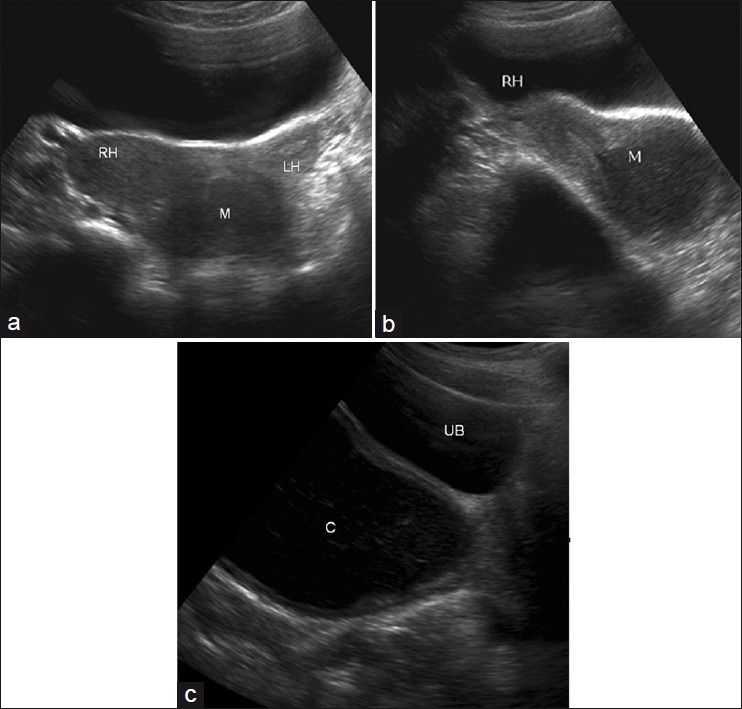
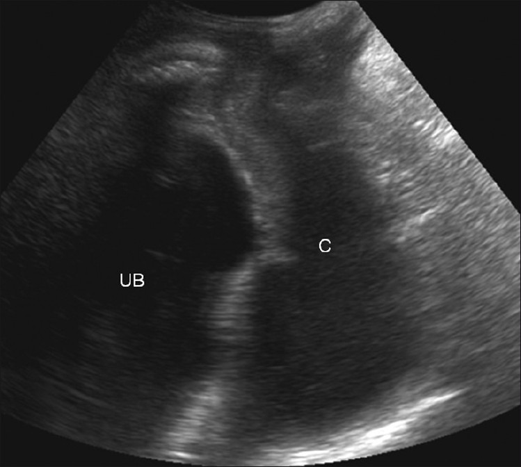
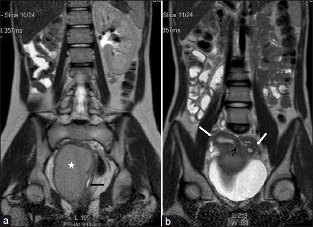
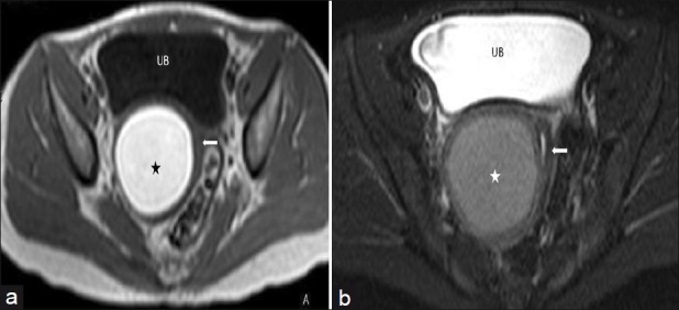
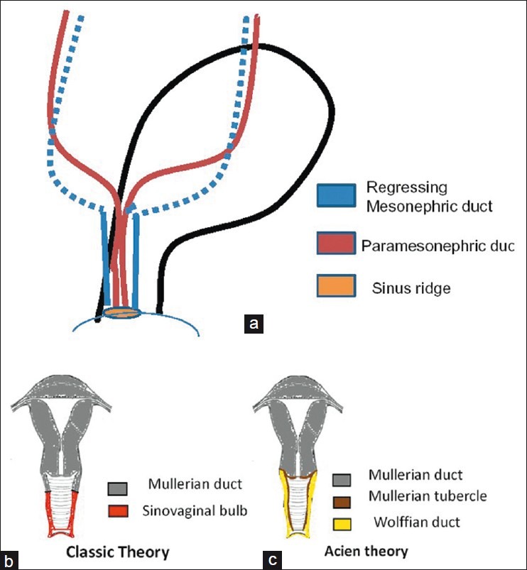
We present here a case of an uncommon complex uterine anomaly - Obstructed HemiVagina with Ipsilateral Renal Agenesis (OHVIRA), also known as Herlyn-Werner-Wunderlich syndrome in a 14-year-old girl along with sonographic (trans-abdominal and trans labial), and MRI findings. The patient underwent surgery wherein imaging findings were confirmed. An MRI has proved to be of great help in correct diagnosis avoiding surgical interventions/ laparoscopy, which were needed in past to diagnose this rare anomaly. We also discuss the development of this anomaly with the help of a relatively new theory of uro-genital development by Acien and review the literature.

Keywords: Herlyn-Werner-Wunderlich syndrome; OHVIRA syndrome; mullerian anomaly; obstructed vagina.


Conflict of interest statement

Conflict of Interest: None declared.

Figures

Figure 1
Figure 1
(a) Axial trans abdominal USG image showing right (RH) and left (LH) uterine horns. A hypoechoic lesion (M) is seen caudally (b) Sagittal trans abdominal USG image showing endometrial cavity of the right uterine horn (RH) communicating with the hypoechoic lesion (M) (c) Sagittal USG image directed further caudally shows the hypoechoic lesion to be an ovoid fluid collection with internal echoes, located posterior to the urinary bladder. It appears to end slightly above the introitus
Figure 2
Figure 2
Trans labial sagittal USG image confirms that the collection (C) ends 1.5 cm above the introitus
Figure 3
Figure 3
(a) Coronal Single shot T2W image shows absence of the right kidney. The distended hemivagina (asterisk) is seen on the right side and the normal collapsed left hemivagina with minimal fluid is seen adjacent to it (black arrow). The distended hemivagina ends above the introitus and its contents are hypointense to fat. (b) Coronal Single shot T2W image shows right and left uterine horns (white arrows). The right uterine horn cavity is seen to communicate with the upper end of the fluid collection in right hemivagina (small black arrow)
Figure 4
Figure 4
(a) Axial T1W image of the pelvis shows bright signal intensity of the right hemivaginal collection (asterisk). The collapsed left hemivagina is seen adjacent to it (arrow) (b) Axial fat saturated T2W image of the pelvis showing right hemivaginal collection (asterisk) and collapsed left hemivagina with minimal fluid (arrow)
Figure 5
Figure 5
Diagrammatic representation of anatomy of patient showing OHVIRA syndrome
Figure 6
Figure 6
(a) Intraoperative photograph showing smooth bulge (black arrow) in medial wall of patent left hemivagina caused by right hematohemicolpos (b) Intraoperative photograph of septoplasty procedure showing drainage of altered blood
Figure 7
Figure 7
(a) Embryological development of the female urogenital system (b) Classic theory of development of uterus and vagina (c) Acien theory of development of uterus and vagina

References

    1. Strassmann EO. Fertility and unification of double uterus. Fertil Steril. 1966;17:165–76. - PubMed
    1. Acien P. Incidence of Müllerian defects in fertile and infertile women. Hum Reprod. 1997;12:1372–6. - PubMed
    1. Li S, Qayyum A, Coakley FV, Hricak H. Association of renal agenesis and mullerian duct anomalies. J Comput Assist Tomogr. 2000;24:829–34. - PubMed
    1. Acién P. Embryological observations on the female genital tract. Hum Reprod. 1992;7:437–45. - PubMed
    1. Koff AK. Development of the vagina in the human fetus. Contrib Embryol. 1933;24:59–91. - PubMed

Publication types