

HSP27 modulates epithelial to mesenchymal transition of lung cancer cells in a Smad-independent manner
- PMID: 22870103
- PMCID: PMC3412513
- DOI: 10.3892/ol.2010.190
HSP27 modulates epithelial to mesenchymal transition of lung cancer cells in a Smad-independent manner
Abstract
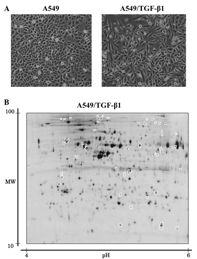
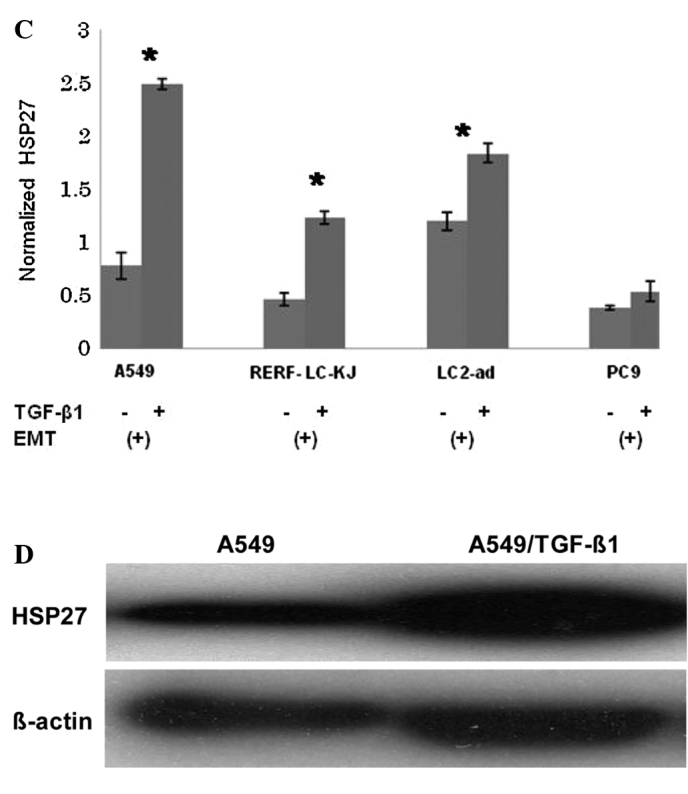
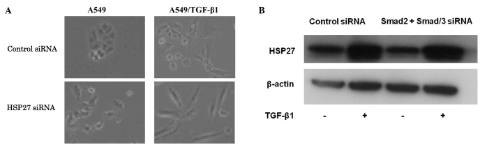
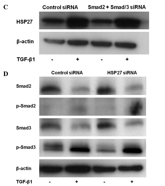
Epithelial to mesenchymal transition (EMT) is induced by transforming growth factor-β1 (TGF-β1) and is a crucial event for cancer cells to acquire invasive and metastatic phenotypes. However, the signals that induce EMT in cancer cells have yet to be adequately defined. In this study, a proteomic investigation was performed to understand the signaling pathway of the EMT of lung cancer using two-dimensional difference gel electrophoresis (2D-DIGE) and mass spectrometry. The protein expression profiles of A549 were compared to those of A549 cells treated with TGF-β1. Of more than 2,000 protein spots shown by 2D-DIGE, 53 were found to be up- or down-regulated upon induction with TGF-β1. In the 53 protein spots, the protein level of heat shock protein (HSP) 27 was found to increase significantly. HSP27 protein was higher in two different lung cancer cell lines, demonstrating the EMT phenomenon with TGF-β1. Notably, the silencing of HSP27 enhanced spindle integration, resulting in an additive effect with TGF-β1-induced EMT. Furthermore, the TGF-β1-induced HSP27 increase was not affected by the suppression of Smad2 and Smad3 in A549 cells. These results suggest that HSP27 was involved in TGF-β1-induced EMT in a Smad-independent manner in lung cancer cells and may provide an effective clinical strategy in lung cancer patients whose tumors are dependent on TGF-β1-induced EMT.
Figures
Similar articles
-
Exogenous hydrogen sulfide donor NaHS alleviates nickel-induced epithelial-mesenchymal transition and the migration of A549 cells by regulating TGF-β1/Smad2/Smad3 signaling.Ecotoxicol Environ Saf. 2020 Jun 1;195:110464. doi: 10.1016/j.ecoenv.2020.110464. Epub 2020 Mar 12. Ecotoxicol Environ Saf. 2020. PMID: 32171946
-
Calreticulin regulates TGF-β1-induced epithelial mesenchymal transition through modulating Smad signaling and calcium signaling.Int J Biochem Cell Biol. 2017 Sep;90:103-113. doi: 10.1016/j.biocel.2017.07.023. Epub 2017 Aug 1. Int J Biochem Cell Biol. 2017. PMID: 28778674
-
Chinese medicine Bu-Fei decoction attenuates epithelial-mesenchymal transition of non-small cell lung cancer via inhibition of transforming growth factor β1 signaling pathway in vitro and in vivo.J Ethnopharmacol. 2017 May 23;204:45-57. doi: 10.1016/j.jep.2017.04.008. Epub 2017 Apr 12. J Ethnopharmacol. 2017. PMID: 28412214
-
TGF-beta1 induces human alveolar epithelial to mesenchymal cell transition (EMT).Respir Res. 2005 Jun 9;6(1):56. doi: 10.1186/1465-9921-6-56. Respir Res. 2005. PMID: 15946381 Free PMC article.
-
Sanguiin H6 suppresses TGF-β induction of the epithelial-mesenchymal transition and inhibits migration and invasion in A549 lung cancer.Bioorg Med Chem Lett. 2015 Dec 1;25(23):5508-13. doi: 10.1016/j.bmcl.2015.10.067. Epub 2015 Oct 23. Bioorg Med Chem Lett. 2015. PMID: 26508552
Cited by
-
HSP27 associates with epithelial-mesenchymal transition, stemness and radioresistance of salivary adenoid cystic carcinoma.J Cell Mol Med. 2018 Apr;22(4):2283-2298. doi: 10.1111/jcmm.13510. Epub 2018 Feb 9. J Cell Mol Med. 2018. Retraction in: J Cell Mol Med. 2025 Jan;29(1):e70365. doi: 10.1111/jcmm.70365. PMID: 29424489 Free PMC article. Retracted.
-
Research on the efficacy of Celastrus Orbiculatus in suppressing TGF-β1-induced epithelial-mesenchymal transition by inhibiting HSP27 and TNF-α-induced NF-κ B/Snail signaling pathway in human gastric adenocarcinoma.BMC Complement Altern Med. 2014 Nov 5;14:433. doi: 10.1186/1472-6882-14-433. BMC Complement Altern Med. 2014. Retraction in: BMC Complement Med Ther. 2025 Jan 7;25(1):2. doi: 10.1186/s12906-024-04741-6. PMID: 25370696 Free PMC article. Retracted.
-
HSP27/Menin Expression as New Prognostic Serum Biomarkers of Prostate Cancer Aggressiveness Independent of PSA.Cancers (Basel). 2022 Sep 29;14(19):4773. doi: 10.3390/cancers14194773. Cancers (Basel). 2022. PMID: 36230697 Free PMC article.
-
Inactivation of p38 MAPK contributes to stem cell-like properties of non-small cell lung cancer.Oncotarget. 2017 Apr 18;8(16):26702-26717. doi: 10.18632/oncotarget.15804. Oncotarget. 2017. PMID: 28460458 Free PMC article.
-
Long noncoding RNA BX357664 regulates cell proliferation and epithelial-to-mesenchymal transition via inhibition of TGF-β1/p38/HSP27 signaling in renal cell carcinoma.Oncotarget. 2016 Dec 6;7(49):81410-81422. doi: 10.18632/oncotarget.12937. Oncotarget. 2016. PMID: 27806310 Free PMC article.
References
-
- Grünert S, Jechlinger M, Beug H. Diverse cellular and molecular mechanisms contribute to epithelial plasticity and metastasis. Nat Rev Mol Cell Biol. 2003;4:657–665. - PubMed
-
- Thiery JP. Epithelial-mesenchymal transitions in development and pathologies. Curr Opin Cell Biol. 2003;15:740–746. - PubMed
-
- Thiery JP. Epithelial-mesenchymal transitions in tumour progression. Nat Rev Cancer. 2002;2:442–454. - PubMed
-
- Huber MA, Kraut N, Beug H. Molecular requirements for epithelial-mesenchymal transition during tumor progression. Curr Opin Cell Biol. 2005;17:548–558. - PubMed
LinkOut - more resources
Full Text Sources
Research Materials
Miscellaneous