. 2010 Nov;1(6):1061-1066.
doi: 10.3892/ol.2010.171. Epub 2010 Sep 9.

Expression of α2,6-sialic acid-containing and Lewis-active glycolipids in several types of human ovarian carcinomas

Affiliations

Expression of α2,6-sialic acid-containing and Lewis-active glycolipids in several types of human ovarian carcinomas

Kyoko Tanaka et al. Oncol Lett. 2010 Nov.

Abstract

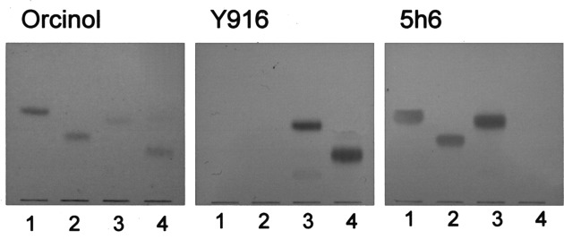
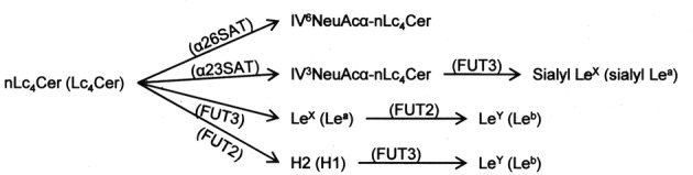
To identify glycolipid antigens associated with histologically defined types of ovarian carcinomas, we determined the amounts of α2,6-sialyl and Lewis-active glycolipids, the specific activities of the α2,3- and α2,6-sialyltransferases, and the gene expression of sugar transferases in mucinous and serous cystadenocarcinoma, clear cell adenocarcinoma and endometrioid carcinoma tissues and cell lines derived from them. α2,6-sialyl glycolipid IV(6)NeuAcα-nLc(4)Cer detected with a newly developed monoclonal antibody, Y916, was present in 5/7 serous cystadenocarcinoma cases in relatively higher amounts than those in the other carcinoma tissues. On the other hand, the amounts of Lewis-active glycolipids in serous cystadenocarcinoma tissues were lower than those in the other carcinoma tissues. No correlation was observed between the structures of Lewis glycolipids and the histological classification. The gene expression of α2,3- and α2,6-sialyltransferases and α1,3/4-fucosyltransferase for the synthesis of Lewis-active glycolipids was not positively correlated with the amounts of the respective glycolipids, probably due to the epigenetic regulation of transferases in the overall metabolic pathways for lacto-series glycolipids. However, the amounts of GM3 and GD3 with short carbohydrate chains correlated with the relative intensities of GM3 and GD3 synthase gene expression, respectively. Among ovarian carcinoma-derived cell lines, the serous cystadenocarcinoma-derived ones exhibited a lower frequency of Lewis-active glycolipid expression than the other carcinoma-derived ones, which was similar to that in the respective tissues. Thus, malignancy-related Lewis-active glycolipids were shown to be regulated in different modes in ovarian serous cystadenocarcinomas and the other carcinomas.


Figures

Figure 1
Figure 1
Thin layer chromatography (TLC) and TLC-immunostaining of glycolipids with monoclonal antibodies Y916 and 5h6. (1) GM3, (2) IV3NeuAcα-nLc4Cer, (3) GD3 and (4) IV6NeuAcα-nLc4Cer were developed on plastic-coated TLC plates with chloroform/methanol/0.5% CaCl2 in water (55:45:10, by volume). Spots were visualized with orcinol-H2SO4 reagent and monoclonal antibodies Y916 and 5h6.
Figure 2
Figure 2
TLC-immunostaining of glycolipids from human ovarian carcinoma tissues. Glycolipids, corresponding to 0.2 mg dry weight, were developed with chloroform/methanol/0.5% CaCl2 in water (55:45:10, by volume). Spots were visualized by TLC-immunostaining with anti-Leb, anti-LeY, anti-LeX, anti-sialyl Lea, and Y916 and 5h6 antibodies. The specimen numbers are the same as those shown in Table I. Standard glycolipids for Y916 was a mixture of GD3 and IV6NeuAcα-nLc4Cer.
Figure 3
Figure 3
TLC-immunostaining of glycolipids from human ovarian mucinous and serous cystadenocarcinoma tissues. Glycolipids were obtained from an additional five specimens, respectively, i.e., other than those in Fig. 2 and Table I. The glycolipids, corresponding to 0.1 mg dry weight, were used for TLC-immunostaining with monoclonal Y916 and anti-Leb antibodies as described in Materials and methods. Standard glycolipids for Y916 was a mixture of GD3 and IV6NeuAcα-nLc4Cer.
Figure 4
Figure 4
TLC-immunostaining of glycolipids from ovarian carcinoma-derived cells. Glycolipids, corresponding to 0.2 mg dry weight, were used for TLC-immunostaining with anti-Leb and anti-LeY antibodies. Lanes: 1, RMG-1; 2, HAC-2; 3, HMKOA; 4, HNOA; 5, 2008 and 6, KF28.
Figure 5
Figure 5
TLC-immunostaining of products following reactions of α2,3- and α2,6-sialyltransferases (SAT). The products following the reactions with 50 μg of enzyme proteins were developed with chloroform/methanol/0.5% CaCl2 in water (55:45:10, by volume), and detected by TLC-immunostaining with monoclonal antibodies 5h6 for IV3NeuAcα-nLc4Cer (α2,3SAT) and Y916 for IV6NeuAcα-nLc4Cer (α2,6SAT), respectively. Standard glycolipids IV3NeuAcα-nLc4Cer for α2,3SAT and IV6NeuAcα-nLc4Cer for α2,6SAT, respectively. The specimen numbers are the same as those in Fig. 2 and Table I.
Figure 6
Figure 6
RT-PCR analysis of the glycosyltransferase genes in human ovarian carcinoma tissues. A, GAPDH; B, GM3 synthase; C, GD3 synthase; D, α2,3-sialyltransferase; E, α2,6-sialyltransferase and F, α1,3/4-fucosyltransferase 3. The specimen numbers are the same as those in Fig. 2 and Table I.
Figure 7
Figure 7
Metabolic pathways for the syntheses of IV6NeuAcα-nLc4Cer and Lewis-active glycolipids. SAT, sialyltransferase; FUT2, α1,2-fucosyltrans-ferase; FUT3, α1,3/4-fucosyltransferase; H, blood group H antigen.

Similar articles

Cited by

References

    1. Roseman S. Reflections on glycobiology. J Biol Chem. 2001;276:41527–41542. - PubMed
    1. Koprowski H, Herlyn M, Steplewski Z, Sears HF. Specific antigen in serum of patients with colon carcinoma. Science. 1981;212:53–55. - PubMed
    1. Canney PA, Wilkinson PM, James RD, Moore M. CA19-9 as a marker for ovarian cancer: alone and in comparison with CA125. Br J Cancer. 1985;52:131–133. - PMC - PubMed
    1. Sgroi D, Varki A, Braesch-Andersen S, Stamenkovic I. CD22, a B cell-specific immunoglobulin superfamily member, is a sialic acid-binding lectin. J Biol Chem. 1993;268:7011–7018. - PubMed
    1. Springer TA, Lasky LA. Cell adhesion. Sticky sugars for selectins. Nature. 1991;349:196–197. - PubMed

LinkOut - more resources