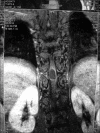
Isolated intramedullary spinal cord cysticercosis
- PMID: 22870160
- PMCID: PMC3410169
- DOI: 10.4103/1793-5482.98655
Isolated intramedullary spinal cord cysticercosis
Abstract
We report a case of intradural, intramedullary, spinal cord neurocysticercosis at dorsal 10-11 (D10-11) level in a mentally retarded male. A 38-year-old, mentally retarded male presented with weakness and stiffness in both the lower limbs and waist since one year. Magnetic resonance imaging revealed a D10-D11 intradural space occupying lesion with cord compression. Intraoperatively, the tumor was grayish white, soft, cystic, and intramedullary with a well-defined plane with surrounding cord tissue. Gross examination revealed a cystic lesion of 1.5×1×0.8 cm, with a whitish nodule of 0.3 cm in diameter. The cyst wall was thin, shiny, and translucent. Microscopic examination revealed cysticercous cyst. Spinal neurocysticercosis should be considered in differential diagnosis of spinal mass lesion in patients residing in endemic area such as India.
Keywords: Cysticercosis; intramedullary; neurocysticercosis; spinal cord.
Conflict of interest statement
Figures
Similar articles
-
Isolated intramedullary spinal cord cysticercosis.J Neurosci Rural Pract. 2014 Nov;5(Suppl 1):S66-8. doi: 10.4103/0976-3147.145209. J Neurosci Rural Pract. 2014. PMID: 25540546 Free PMC article.
-
Intramedullary spinal cysticercosis simulating a conus medullaris tumor: case report.Arq Neuropsiquiatr. 2006 Mar;64(1):149-52. doi: 10.1590/s0004-282x2006000100033. Epub 2006 Apr 5. Arq Neuropsiquiatr. 2006. PMID: 16622575
-
A Rare Case of Isolated Intramedullary Spinal Cord Cysticercosis.Cureus. 2021 May 6;13(5):e14864. doi: 10.7759/cureus.14864. Cureus. 2021. PMID: 34113502 Free PMC article.
-
Spinal intramedullary cysticercosis: a case report and literature review.Int J Med Sci. 2011;8(5):420-3. doi: 10.7150/ijms.8.420. Epub 2011 Jul 6. Int J Med Sci. 2011. PMID: 21814474 Free PMC article. Review.
-
Intramedullary cysticercosis of the spinal cord: a review of patients evaluated with MRI.J Neurol Sci. 2013 Aug 15;331(1-2):114-7. doi: 10.1016/j.jns.2013.05.025. Epub 2013 Jun 6. J Neurol Sci. 2013. PMID: 23747004 Review.
Cited by
-
Spinal intramedullary cysticercosis with syringomyelia: a case report.Int J Clin Exp Pathol. 2020 Oct 1;13(10):2593-2598. eCollection 2020. Int J Clin Exp Pathol. 2020. PMID: 33165438 Free PMC article.
-
Case report and review of literature: Isolated intramedullary spinal neurocysticercosis.Front Neurol. 2022 Nov 10;13:1030468. doi: 10.3389/fneur.2022.1030468. eCollection 2022. Front Neurol. 2022. PMID: 36438967 Free PMC article.
-
Isolated Intramedullary Lumbar Spine Neurocysticercosis: A Rare Occurrence and Review of Literature.Surg J (N Y). 2021 Dec 15;7(4):e327-e336. doi: 10.1055/s-0041-1739118. eCollection 2021 Oct. Surg J (N Y). 2021. PMID: 34926816 Free PMC article.
-
Isolated intramedullary spinal cord cysticercosis.J Neurosci Rural Pract. 2014 Nov;5(Suppl 1):S66-8. doi: 10.4103/0976-3147.145209. J Neurosci Rural Pract. 2014. PMID: 25540546 Free PMC article.
-
Commentary.J Neurosci Rural Pract. 2014 Nov;5(Suppl 1):S68-70. J Neurosci Rural Pract. 2014. PMID: 25540547 Free PMC article. No abstract available.
References
-
- Colli BO, Valenca MM, Carlotti CG, Jr, Machado HR, Assirati JA., Jr Spinal cord cysticercosis: Neurological aspects. Neurosurg Focus. 2002;12:9. - PubMed
-
- Flisser A, Sarti E, Lightowlers M, Schantz P. Neuro cysticercosis: Regional status, epidemiology, impact and control measures in the Americas. Acta Trop. 2003;87:43–51. - PubMed
-
- Rajeshwar V, Joshi DD, Doanh NQ, van De N, Xiaonong Z. Taenia solium taeniosis/cysticercosis in Asia: Epidemiology, impact and issues. Acta Trop. 2003;87:53–60. - PubMed
-
- Mohanty A, Das S, Kolluri VR, Das BS. Spinal extradural cysticercosis: A case report. Spinal Cord. 1998;36:285–7. - PubMed
-
- Alsina GA, Johnson JP, McBridge DQ, Rhoten PR, Mehringer CM, Stokes JK. Spinal neurocysticercosis. Neurosurg Focus. 2002;12:1–7. - PubMed


