

Petrous bone epidermoid cyst caused by penetrating injury to the external ear: Case report and review of literature
- PMID: 22870161
- PMCID: PMC3410170
- DOI: 10.4103/1793-5482.98656
Petrous bone epidermoid cyst caused by penetrating injury to the external ear: Case report and review of literature
Abstract
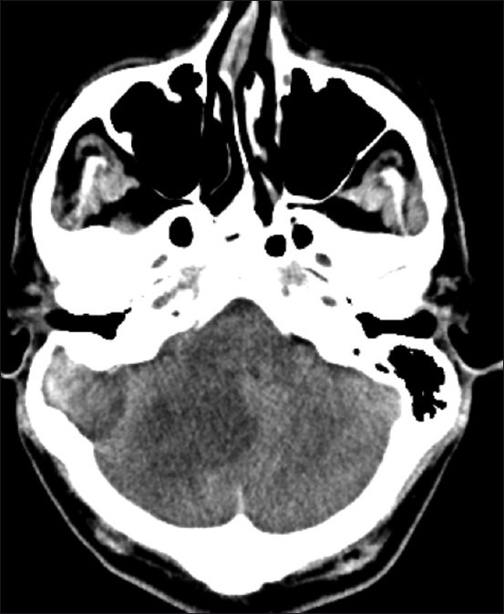
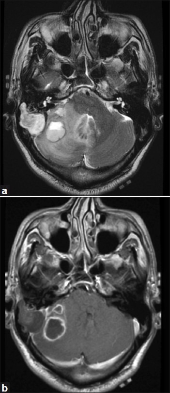
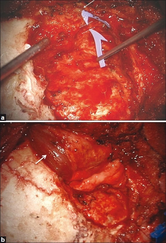
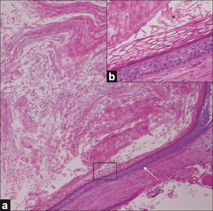
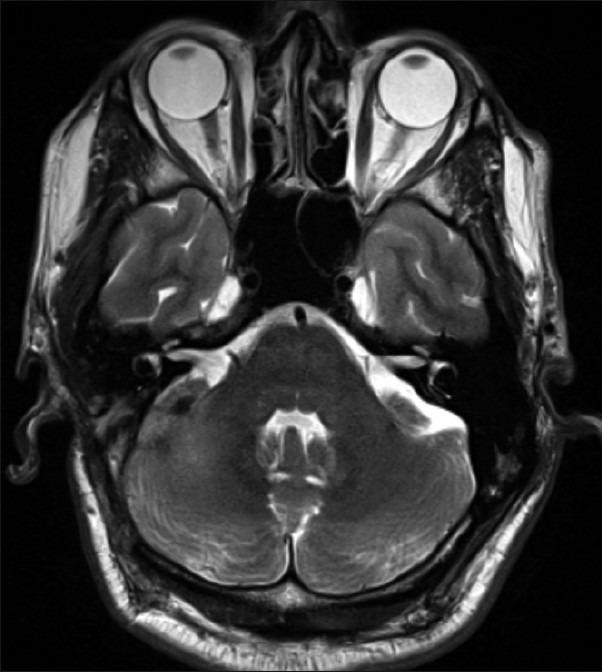
Epidermoid cysts are histologically benign, slow-growing congenital neoplasms of the central nervous system that may arise from retained ectodermal implants. The epidermoid lesions are generally caused during the 3(rd) to 5(th) week of gestation by an incomplete cleavage of the neural tissue from the cutaneous ectoderm, though it can also happen later in life due to introduction of skin elements by skin puncture, trauma or surgery. We present this unique case of a petromastoid epidermoid cyst associated with ipsilateral cerebellar abscesses, presenting 20 years after a penetrating trauma to the external auditory canal. Radical excision of both lesions and revision of the previous fistulous tract was performed. We present the diagnostic challenge and the operative treatment of this unique case, which to our knowledge is the first where an epidermoid cyst and an adjacent brain abscess occurred as a result of a single traumatic event.
Keywords: Epidermoid cyst; penetrating trauma; petrous bone; surgical treatment.
Conflict of interest statement
Figures

References
-
- Hamel E, Frowein RA, Karimi-Nejad A. Intracranial intradural epidermoids and dermoids. Neurosurg Rev. 1980;3:215–9. - PubMed
-
- Revilla AG. Differential diagnosis of tumors at the cerebellopontine recess. Bull John Hopkins Hosp. 1948;83:187–212. - PubMed
-
- Ulrich J. Intracranial epidermoids: A study on their distribution and spread. J Neurosurg. 1964;21:1051–8. - PubMed
-
- Zhou LF. Intracranial epidermoid tumors: Thirty-seven years of diagnosis and treatment. Br J Neurosurg. 1990;4:211–6. - PubMed
-
- Rubin G, Scienza R, Pasqualin A, Rosta L, Da Pian R. Craniocerebral epidermoids and dermoids. Acta Neurochir (Wien) 1989;97:1–16. - PubMed
Publication types
LinkOut - more resources
Full Text Sources
Miscellaneous