. 2012;7(8):e41836.
doi: 10.1371/journal.pone.0041836. Epub 2012 Aug 1.

Carbon monoxide improves cardiac function and mitochondrial population quality in a mouse model of metabolic syndrome

Affiliations

Carbon monoxide improves cardiac function and mitochondrial population quality in a mouse model of metabolic syndrome

Steve Lancel et al. PLoS One. 2012.

Abstract

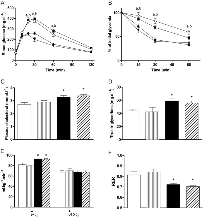
Aims: Metabolic syndrome induces cardiac dysfunction associated with mitochondria abnormalities. As low levels of carbon monoxide (CO) may improve myocardial and mitochondrial activities, we tested whether a CO-releasing molecule (CORM-3) reverses metabolic syndrome-induced cardiac alteration through changes in mitochondrial biogenesis, dynamics and autophagy.

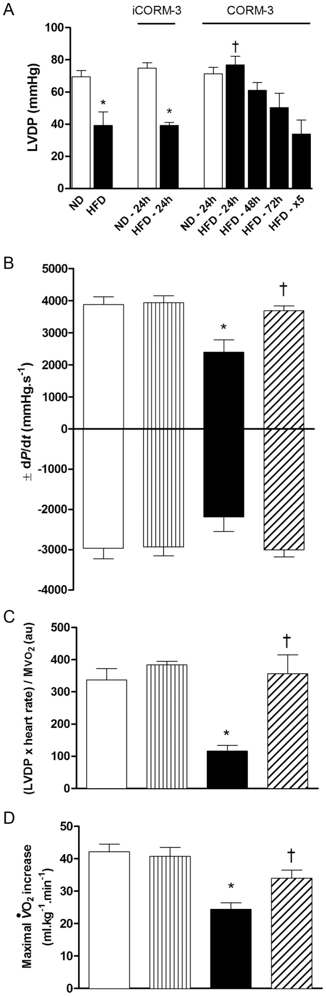
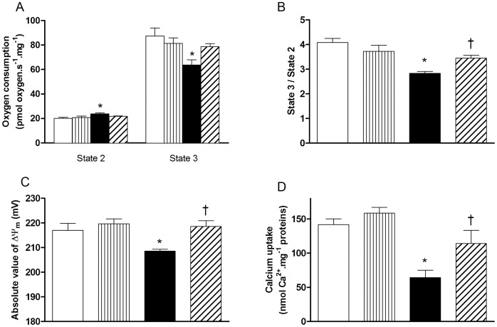
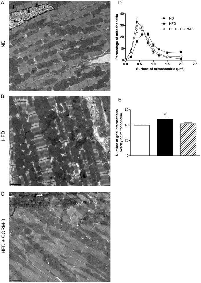
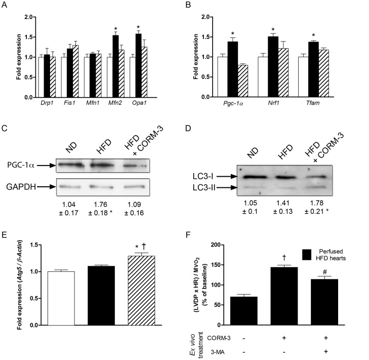
Methods and results: Mice were fed with normal diet (ND) or high-fat diet (HFD) for twelve weeks. Then, mice received two intraperitoneal injections of CORM-3 (10 mg x kg(-1)), with the second one given 16 hours after the first. Contractile function in isolated hearts and mitochondrial parameters were evaluated 24 hours after the last injection. Mitochondrial population was explored by electron microscopy. Changes in mitochondrial dynamics, biogenesis and autophagy were assessed by western-blot and RT-qPCR. Left ventricular developed pressure was reduced in HFD hearts. Mitochondria from HFD hearts presented reduced membrane potential and diminished ADP-coupled respiration. CORM-3 restored both cardiac and mitochondrial functions. Size and number of mitochondria increased in the HFD hearts but not in the CORM-3-treated HFD group. CORM-3 modulated HFD-activated mitochondrial fusion and biogenesis signalling. While autophagy was not activated in the HFD group, CORM-3 increased the autophagy marker LC3-II. Finally, ex vivo experiments demonstrated that autophagy inhibition by 3-methyladenine abolished the cardioprotective effects of CORM-3.

Conclusion: CORM-3 may modulate pathways controlling mitochondrial quality, thus leading to improvements of mitochondrial efficiency and HFD-induced cardiac dysfunction.


Conflict of interest statement

Competing Interests: The authors have declared that no competing interests exist.

Figures

Figure 1
Figure 1. Effects of CORM-3 on high-fat-induced cardiac dysfunction.
(A) Left Ventricular Developed Pressure (LVDP) of hearts isolated from normal diet (ND) or high-fat diet (HFD) mice injected twice with iCORM-3 (10 mg.kg−1, intraperitoneally, studied 24 hours after the second injection) or CORM-3 (10 mg.kg−1, intraperitoneally, studied 24, 48 or 72 hours after the second injection, or 24 hours after the fifth injection (x5); see Materials and Methods). (B) LVDP first derivatives ±dP/dt and (C) ratio (LVDP x Heart Rate)/MVO2 (see Materials and Methods) obtained from ND, ND + CORM-3, HFD and CORM-3–treated HFD mouse hearts. Evaluation was performed 24 hours after the second injection. (D) Maximal oxygen uptake (VO2) increase of mice subjected to exercise stress test. Data represent means ± SEM. n  = 5–8 in each group. White bars  =  ND; vertically hatched bars  =  ND + CORM-3; black bars  =  HFD; crosshatched bars  =  HFD + CORM-3. * p<0.05 vs. ND, † p<0.05 vs. HFD.
Figure 2
Figure 2. Effects of CORM-3 on metabolic parameters.
(A) Oral glucose tolerance and (B) insulin tolerance tests were performed on ND (black circles), ND + CORM-3 (black triangles), HFD (black squares) and HFD + CORM-3 (white diamonds) mice. Data are means ± SEM. n  = 10 in each group. (a) p<0.05 between HFD and ND; (b) p<0.05 between HFD + CORM-3 and ND. (C) Plasma cholesterol and (D) true triglyceride measurements were performed on 6–10 different samples in each group. Data are means ± SEM. (E) Basal oxygen uptake (VO2) and carbon dioxide rejection (VCO2), (F) respiratory exchange ratio (RER). Data represent means ± SEM. n  = 5–7 in each group, * p<0.05 vs. ND. White bars  =  ND; vertically hatched bars  =  ND + CORM-3; black bars  =  HFD; crosshatched bars  =  HFD + CORM-3.
Figure 3
Figure 3. CORM-3 improved mitochondrial function.
(A) Oxygen consumption of permeabilized fibers in presence of glutamate and malate without exogenous ADP (state 2) or with 2.5 mM ADP (state 3) and (B) state 3 to state 2 ratio. Mitochondrial membrane potential (C) and calcium retention capacity (D) measured on isolated cardiac mitochondria. White bars  =  ND; vertically hatched bars  =  ND + CORM-3; black bars  =  HFD; crosshatched bars  =  CORM-3–treated HFD mice. Data are means ± SEM. n  = 6–8 in each group. * p<0.05 vs. ND, † p<0.05 vs. HFD.
Figure 4
Figure 4. CORM-3 did not change UCP expression.
(A) Results of quantitative RT-PCR detecting Ucp2 and Ucp3 mRNA expressions. Results were normalized to â-actin. (B) Representative western-blot targeting UCP3 and GAPDH proteins and (C) densitometric analysis of UCP3 to GAPDH ratio. White bars  =  ND; vertically hatched bars  =  ND + CORM-3; black bars  =  HFD; crosshatched bars  =  CORM-3–treated HFD mice. Data are means ± SEM. n  = 5 in each group. * p<0.05 vs. ND.
Figure 5
Figure 5. CORM-3 modified cardiac mitochondrial population.
(A) Representative electron micrographs of ND, HFD and HFD + CORM-3 mouse hearts. Magnification x7000, scale bar  = 2 µm. (B) Surface of every single mitochondrion from three randomized tissue sections prepared from four hearts in each group was measured. Black circles: ND; black squares: HFD; white diamonds: HFD + CORM-3. Results are expressed in percentage. (C) A grid of 144 µm2 was positioned onto each micrograph (3 sections per heart, 4 hearts per group) and the number of intersections with mitochondria was scored. White bars  =  ND; black bars  =  HFD; crosshatched bars  =  CORM-3–treated HFD mice. Data are means ± SEM. * p<0.05 vs. ND.
Figure 6
Figure 6. Effects of CORM-3 on pathways controlling mitochondrial quality.
RT-qPCR results of fusion/fission (A) and biogenesis (B) -related transcripts in cardiac tissue obtained from ND (white bars), HFD (black bars) or HFD + CORM-3 (crosshatched bars) mice. Data were normalized to â-actin. (C) Representative western-blot of PGC-1α and GAPDH in ND, HFD and HFD + CORM-3 mouse hearts. Values are results of densitometric analysis normalized to GAPDH. (D) Representative western-blot of LC3 proteins in ND, HFD and HFD + CORM-3 mouse hearts. LC3-I represents the unactivated form; LC-II is the activated protein that has been lipidated. Numbers represent the pro-autophagy ratio LC3-II to LC3-I obtained after densitometric analysis. (E) Atg5 RT-qPCR results normalized to â-actin. White bars  =  ND; black bars  =  HFD; crosshatched bars  =  CORM-3–treated HFD mice. Data are means ± SEM. n  = 5 in each group. * p<0.05 vs. ND, † p<0.05 vs. HFD. (F) Ratio (LVDP x Heart Rate)/MVO2 of isolated perfused heart from HFD mice. HFD hearts were pretreated or not with 10 mM 3-methyladenine (3-MA) for 15 min and then perfused with Krebs-Henseleit buffer containing 10 µM of CORM-3. Measurement was performed 60 min after CORM-3 perfusion. Black bars: isolated and perfused HFD hearts. Data are means ± SEM. n  = 4–5 in each group. † p<0.05 vs. untreated HFD hearts, # p<0.05 vs. CORM-3 treated HFD hearts.

References

    1. Boudina S, Abel ED (2010) Diabetic cardiomyopathy, causes and effects. Rev Endocr Metab Disord 11: 31–9. - PMC - PubMed
    1. Ong S-B, Subrayan S, Lim SY, Yellon DM, Davidson SM, et al. (2010) Inhibiting mitochondrial fission protects the heart against ischemia/reperfusion injury. Circulation 121: 2012–22. - PubMed
    1. Lehman JJ, Barger PM, Kovacs A, Saffitz JE, Medeiros DM, et al. (2000) Peroxisome proliferator-activated receptor gamma coactivator-1 promotes cardiac mitochondrial biogenesis. J Clin Invest 106: 847–56. - PMC - PubMed
    1. Nakai A, Yamaguchi O, Takeda T, Higuchi Y, Hikoso S, et al. (2007) The role of autophagy in cardiomyocytes in the basal state and in response to hemodynamic stress. Nat Med 13: 619–24. - PubMed
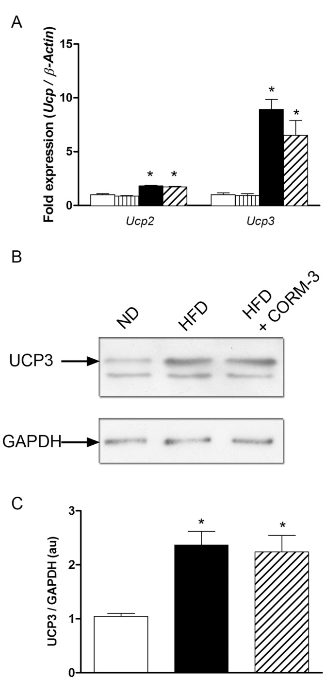
    1. Boudina S, Sena S, Theobald H, Sheng X, Wright JJ, et al. (2007) Mitochondrial energetics in the heart in obesity-related diabetes: direct evidence for increased uncoupled respiration and activation of uncoupling proteins. Diabetes 56: 2457–66. - PubMed

Publication types

MeSH terms