. 2012;7(8):e42863.
doi: 10.1371/journal.pone.0042863. Epub 2012 Aug 3.

The proteolytic activity of separase in BCR-ABL-positive cells is increased by imatinib

Affiliations

The proteolytic activity of separase in BCR-ABL-positive cells is increased by imatinib

Wiltrud Haaß et al. PLoS One. 2012.

Abstract

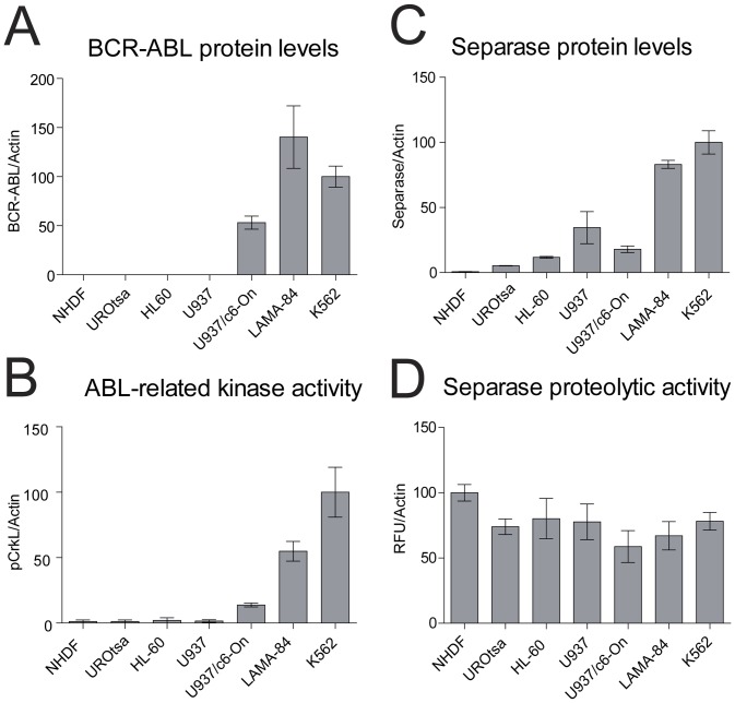
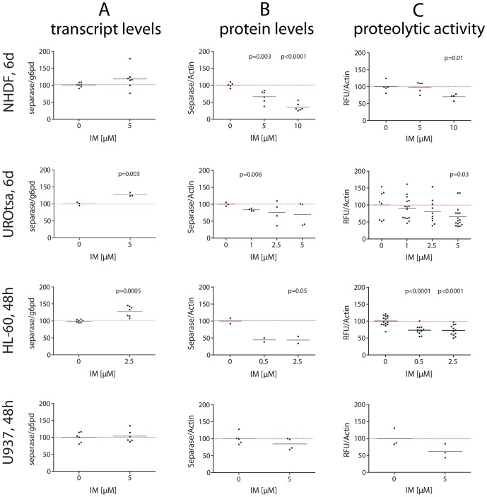
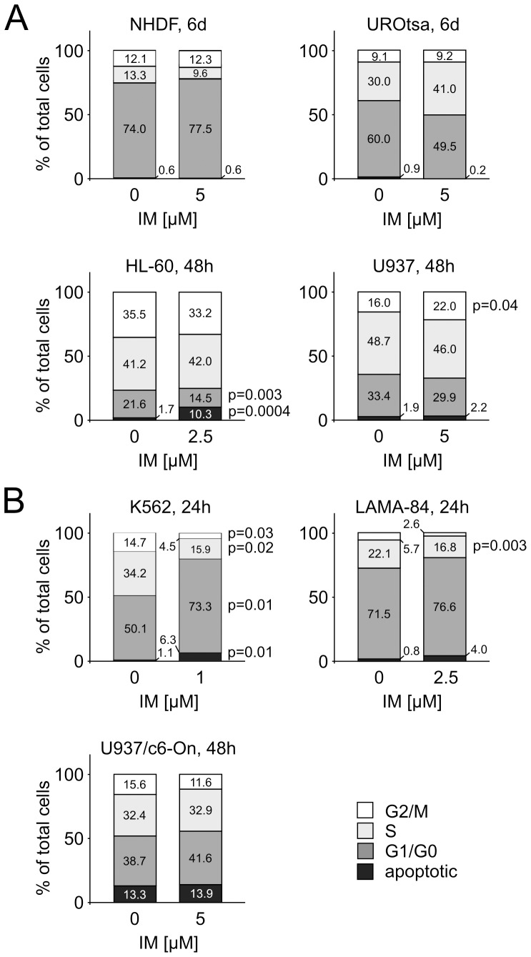
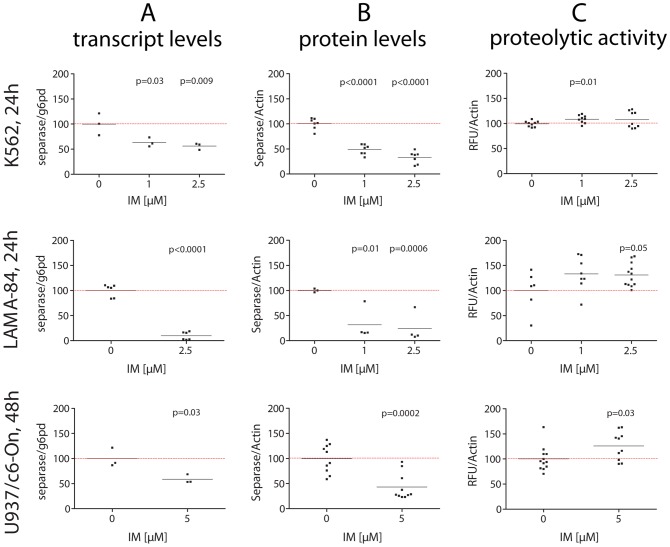
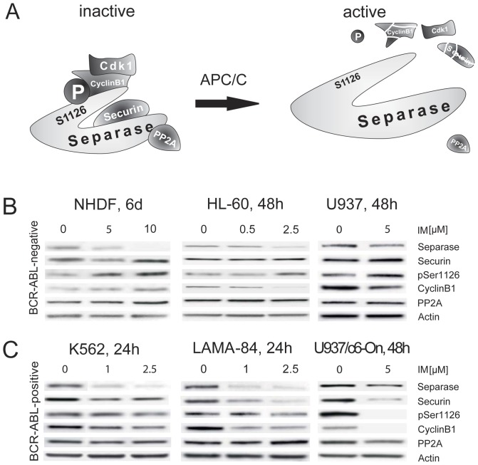
Separase, an endopeptidase required for the separation of sister-chromatides in mitotic anaphase, triggers centriole disengagement during centrosome duplication. In cancer, separase is frequently overexpressed, pointing to a functional role as an aneuploidy promoter associated with centrosomal amplification and genomic instability. Recently, we have shown that centrosomal amplification and subsequent chromosomal aberrations are a hallmark of chronic myeloid leukemia (CML), increasing from chronic phase (CP) toward blast crisis (BC). Moreover, a functional linkage of p210BCR-ABL tyrosine kinase activity with centrosomal amplification and clonal evolution has been established in long-term cell culture experiments. Unexpectedly, therapeutic doses of imatinib (IM) did not counteract; instead induced similar centrosomal alterations in vitro. We investigated the influence of IM and p210BCR-ABL on Separase as a potential driver of centrosomal amplification in CML. Short-term cell cultures of p210BCR-ABL-negative (NHDF, UROtsa, HL-60, U937), positive (K562, LAMA-84) and inducible (U937p210BCR-ABL/c6 (Tet-ON)) human cell lines were treated with therapeutic doses of IM and analyzed by qRT-PCR, Western blot analysis and quantitative Separase activity assays. Decreased Separase protein levels were observed in all cells treated with IM in a dose dependent manner. Accordingly, in all p210BCR-ABL-negative cell lines, decreased proteolytic activity of Separase was found. In contrast, p210BCR-ABL-positive cells showed increased Separase proteolytic activity. This activation of Separase was consistent with changes in the expression levels of Separase regulators (Separase phosphorylation at serine residue 1126, Securin, CyclinB1 and PP2A). Our data suggest that regulation of Separase in IM-treated BCR-ABL-positive cells occurs on both the protein expression and the proteolytic activity levels. Activation of Separase proteolytic activity exclusively in p210BCR-ABL-positive cells during IM treatment may act as a driving force for centrosomal amplification, contributing to genomic instability, clonal evolution and resistance in CML.


Conflict of interest statement

Competing Interests: The authors have declared that no competing interests exist.

Figures

Figure 1
Figure 1. Protein and activity levels of BCR-ABL and Separase in cell lines under investigation.
Protein levels of p210BCR-ABL (A) and Separase (C) based on densitometric evaluation of immunostained Western blots were normalized to Actin as loading control. Abl-related TK activity (BCR-ABL+c-ABL) was measured as pCrkL/Actin (B). Separase proteolytic activity (D) was quantified using an in vitro fluorometric assay and was given as relative fluorescence units/Actin (RFU/Actin). Analyses were performed on protein lysates derived from p210BCR-ABL-positive (LAMA-84, K562) and -negative cells (NHDF, UROtsa, HL-60, U937), and from induced U937p210BCR-ABL/c6 cells (U937/c6-On) expressing a p210BCR-ABL transgene under control of a Doxycycline inducible promoter.
Figure 2
Figure 2. Transcript levels, protein levels and proteolytic activity of Separase in BCR-ABL-negative cells treated with IM.
Cells were treated individually with distinct concentrations (0.5 to 10 µM) of IM. After about two cell cycle rounds (NHDF, UROtsa, 6 d; HL-60 and U937, 48 h) total RNA and protein lysates were prepared and analyzed by qRT-PCR (A), Western blot immunostaining (B) and Separase fluorometric activity assays (C). For Westren blot experiments, Actin served as loading control and/or for densitometric data normalization. Each data point corresponds to one single experiment. Only significant p-values as calculated between treated and untreated cells were shown (see Table 2 for summarized Δ-values). For a representative set of corresponding immunostained Western blots compare Figure 5 panel B.
Figure 3
Figure 3. Cell cycle profiles of untreated and IM-treated cell lines.
Triplicate cell cultures were incubated with IM at times and doses where significant changes in Separase protein levels and/or Separase proteolytic activation have been observed (Figure 2 and 4). Subsequently, the cell cycle profile was analyzed by flow cytometry after propidium iodide staining. The percentages of cells in G2/M, S, G1/G0 phases and in apoptotic state are depicted in each bar.
Figure 4
Figure 4. Transcript levels, protein levels and proteolytic activity of Separase in BCR-ABL-positive cells treated with IM.
Cells were treated individually with distinct concentrations (0.5 to 5 µM) of IM. After about two cell cycle rounds (K562, LAMA-84, 24 h; U937p210BCR-ABL/c6-On, 48 h) total RNA and protein lysates were prepared and analyzed by qRT-PCR (A), Western blot immunostaining (B) and separase fluorometric activity assays (C). For Western blot experiments, Actin served as loading control and/or for densitometric data normalization. Each data point corresponds to one single experiment. Only significant p-values as calculated between treated and untreated cells were shown (see Table 2 for summarized Δ-values). For a representative set of corresponding immunostained Western blots compare Figure 5 panel C. Abbreviations: U937/c6-On, U937 cells expressing a p210BCR-ABL transgene (Tet-On system) after induction with Doxycycline (U937p210BCR-ABL/c6-On).
Figure 5
Figure 5. Analysis of master Separase proteolytic activity regulators in BCR-ABL-negative and -positive cell lines treated with IM.
Schematic diagram of cooperating inhibitory factors that regulate Separase proteolytic activity in a tight cell cycle controlled manner (A). Degradation of Securin, inactivation of the CyclinB1/CDK1 complex, dephosphorylation of Separase at a specific serine residue (pSer1126) by the anaphase promoting complex/cyclosome (APC/C), and the release of PP2A contribute to activation of Separase. Representative composite image of Western blot immunostaining experiments illustrate the expression levels of Separase and relevant regulatory proteins (Securin, pSer1126, CyclinB1 and PP2A) in BCR-ABL-negative (B) and BCR-ABL-positive (C) cell lines. Images are cropped sections derived from stripped and reprobed Western blot immunostainings used for acquisition of densitometric data shown in Figures 2 and 4. Cells were treated with IM for times and doses given on top. Actin served as loading control. The densitometric data of at least triplicate experiments are presented in Table 3.

References

    1. Sawyers CL (1999) Chronic myeloid leukemia. N Engl J Med 340: 1330–1340. - PubMed
    1. Calabretta B, Perrotti D (2004) The biology of CML blast crisis. Blood 103: 4010–4022. - PubMed
    1. Kumari A, Brendel C, Hochhaus A, Neubauer A, Burchert A (2011) Low BCR-ABL expression levels in hematopoietic precursor cells enable persistence of chronic myeloid leukemia under imatinib. Blood 119: 530–539. - PubMed
    1. Druker BJ, Guilhot F, O'Brien SG, Gathmann I, Kantarjian H, et al. (2006) Five-year follow-up of patients receiving imatinib for chronic myeloid leukemia. N Engl J Med 355: 2408–2417. - PubMed
    1. Hochhaus A, Schenk T, Erben P, Ernst T, La Rosee P, et al. (2009) Cause and management of therapy resistance. Best Pract Res Clin Haematol 22: 367–379. - PubMed

Publication types

MeSH terms