A simple and efficient transient transformation for hybrid aspen (Populus tremula × P. tremuloides)
- PMID: 22871142
- PMCID: PMC3476444
- DOI: 10.1186/1746-4811-8-30
A simple and efficient transient transformation for hybrid aspen (Populus tremula × P. tremuloides)
Abstract
Background: The genus Populus is accepted as a model system for molecular tree biology. To investigate gene functions in Populus spp. trees, generating stable transgenic lines is the common technique for functional genetic studies. However, a limited number of genes have been targeted due to the lengthy transgenic process. Transient transformation assays complementing stable transformation have significant advantages for rapid in vivo assessment of gene function. The aim of this study is to develop a simple and efficient transient transformation for hybrid aspen and to provide its potential applications for functional genomic approaches.
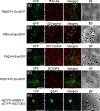
Results: We developed an in planta transient transformation assay for young hybrid aspen cuttings using Agrobacterium-mediated vacuum infiltration. The transformation conditions such as the infiltration medium, the presence of a surfactant, the phase of bacterial growth and bacterial density were optimized to achieve a higher transformation efficiency in young aspen leaves. The Agrobacterium infiltration assay successfully transformed various cell types in leaf tissues. Intracellular localization of four aspen genes was confirmed in homologous Populus spp. using fusion constructs with the green fluorescent protein. Protein-protein interaction was detected in transiently co-transformed cells with bimolecular fluorescence complementation technique. In vivo promoter activity was monitored over a few days in aspen cuttings that were transformed with luciferase reporter gene driven by a circadian clock promoter.
Conclusions: The Agrobacterium infiltration assay developed here is a simple and enhanced throughput method that requires minimum handling and short transgenic process. This method will facilitate functional analyses of Populus genes in a homologous plant system.
Figures


Similar articles
-
An improved and efficient method of Agrobacterium syringe infiltration for transient transformation and its application in the elucidation of gene function in poplar.BMC Plant Biol. 2021 Jan 21;21(1):54. doi: 10.1186/s12870-021-02833-w. BMC Plant Biol. 2021. PMID: 33478390 Free PMC article.
-
An efficient Agrobacterium-mediated transformation and regeneration system for leaf explants of two elite aspen hybrid clones Populus alba × P. berolinensis and Populus davidiana × P. bolleana.Plant Cell Rep. 2011 Nov;30(11):2037-44. doi: 10.1007/s00299-011-1111-1. Epub 2011 Jun 30. Plant Cell Rep. 2011. PMID: 21717184
-
Agrobacterium-mediated transformation of quaking aspen (Populus tremuloides) and regeneration of transgenic plants.Plant Cell Rep. 1994 Dec;14(2-3):94-7. doi: 10.1007/BF00233768. Plant Cell Rep. 1994. PMID: 24192872
-
Targeted integration and removal of transgenes in hybrid aspen (Populus tremula L. x P. tremuloides Michx.) using site-specific recombination systems.Plant Biol (Stuttg). 2010 Mar;12(2):334-40. doi: 10.1111/j.1438-8677.2009.00293.x. Plant Biol (Stuttg). 2010. PMID: 20398239 Review.
-
T-DNA and transposon tagging in aspen.Plant Biol (Stuttg). 2004 Jan-Feb;6(1):5-11. doi: 10.1055/s-2003-44745. Plant Biol (Stuttg). 2004. PMID: 15095129 Review.
Cited by
-
Refinement and Enhancement of Agrobacterium-Mediated Transient Transformation for Functional Gene Examination in Mulberry (Morus L.).Genes (Basel). 2024 Sep 28;15(10):1277. doi: 10.3390/genes15101277. Genes (Basel). 2024. PMID: 39457401 Free PMC article.
-
Discovery of a novel small secreted protein family with conserved N-terminal IGY motif in Dikarya fungi.BMC Genomics. 2014 Dec 20;15(1):1151. doi: 10.1186/1471-2164-15-1151. BMC Genomics. 2014. PMID: 25526808 Free PMC article.
-
A simple and efficient agroinfiltration method in coffee leaves (Coffea arabica L.): assessment of factors affecting transgene expression.3 Biotech. 2018 Nov;8(11):471. doi: 10.1007/s13205-018-1495-5. Epub 2018 Nov 8. 3 Biotech. 2018. PMID: 30456005 Free PMC article.
-
PuHSFA4a Enhances Tolerance To Excess Zinc by Regulating Reactive Oxygen Species Production and Root Development in Populus.Plant Physiol. 2019 Aug;180(4):2254-2271. doi: 10.1104/pp.18.01495. Epub 2019 Jun 20. Plant Physiol. 2019. PMID: 31221731 Free PMC article.
-
A rapid, highly efficient and economical method of Agrobacterium-mediated in planta transient transformation in living onion epidermis.PLoS One. 2014 Jan 8;9(1):e83556. doi: 10.1371/journal.pone.0083556. eCollection 2014. PLoS One. 2014. PMID: 24416168 Free PMC article.
References
-
- Tuskan GA, Difazio S, Jansson S, Bohlmann J, Grigoriev I, Hellsten U, Putnam N, Ralph S, Rombauts S, Salamov A, Schein J, Sterck L, Aerts A, Bhalerao RR, Bhalerao RP, Blaudez D, Boerjan W, Brun A, Brunner A, Busov V, Campbell M, Carlson J, Chalot M, Chapman J, Chen GL, Cooper D, Coutinho PM, Couturier J, Covert S, Cronk Q. et al.The genome of black cottonwood, Populus trichocarpa (Torr. & Gray) Science. 2006;313:1596–1604. doi: 10.1126/science.1128691. - DOI - PubMed
-
- Street NR, Tsai CJ. In: Genetics and Genomics of Populus. Jansson S, Bhalerao R, Groover A, editor. Springer Science+Business Media, LLC, New York; 2010. Populus Resources and Bioinformatics; pp. 135–152.
LinkOut - more resources
Full Text Sources
Miscellaneous

