

Collagen type I and decorin expression in tenocytes depend on the cell isolation method
- PMID: 22871215
- PMCID: PMC3518183
- DOI: 10.1186/1471-2474-13-140
Collagen type I and decorin expression in tenocytes depend on the cell isolation method
Abstract
Background: The treatment of rotator cuff tears is still challenging. Tendon tissue engineering (TTE) might be an alternative in future. Tenocytes seem to be the most suitable cell type as they are easy to obtain and no differentiation in vitro is necessary. The aim of this study was to examine, if the long head of the biceps tendon (LHB) can deliver viable tenocytes for TTE. In this context, different isolation methods, such as enzymatic digestion (ED) and cell migration (CM), are investigated on differences in gene expression and cell morphology.
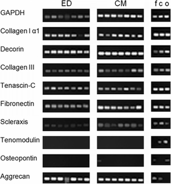
Methods: Samples of the LHB were obtained from patients, who underwent surgery for primary shoulder arthroplasty. Using ED as isolation method, 0.2% collagenase I solution was used. Using CM as isolation method, small pieces of minced tendon were put into petri-dishes. After cell cultivation, RT-PCR was performed for collagen type I, collagen type III, decorin, tenascin-C, fibronectin, Scleraxis, tenomodulin, osteopontin and agreccan.
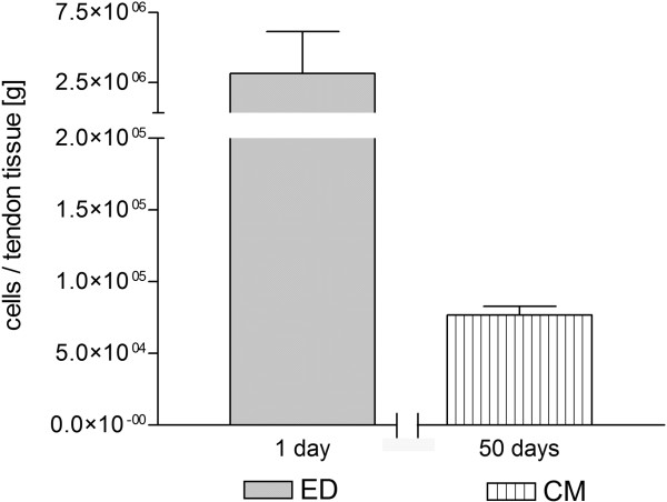
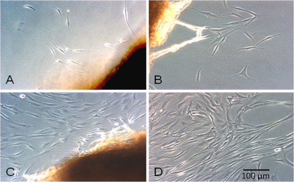
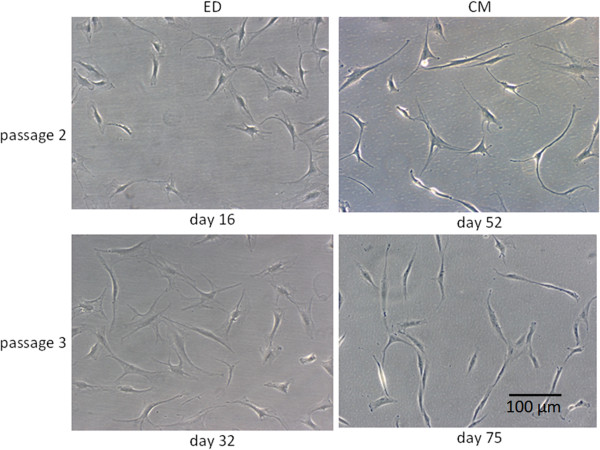
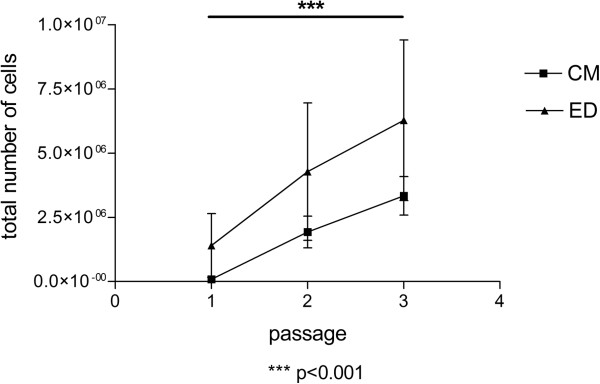
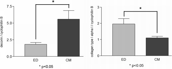
Results: The total number of isolated cells, in relation to 1 g of native tissue, was 14 times higher using ED. The time interval for cell isolation was about 17 hours using ED and approximately 50 days using CM. Cell morphology in vitro was similar for both isolation techniques. Higher expression of collagen type I could be observed in tenocyte-like cell cultures (TLCC) using ED as isolation method (p < 0.05), however decorin expression was higher in TLCC using CM as isolation method (p < 0.05). Dedifferentiation potential seemed to be similar for both isolation techniques.
Conclusion: In summary tenocyte-like cells can be obtained with both isolation methods (ED and CM) from the LHB. As no obvious disadvantage could be seen using ED, this method is more suitable for clinical use, as time for cell isolation is shorter and a remarkably higher number of cells can be obtained. However, both isolation methods can further be improved.
Figures
Similar articles
-
In vitro changes in human tenocyte cultures obtained from proximal biceps tendon: multiple passages result in changes in routine cell markers.Knee Surg Sports Traumatol Arthrosc. 2012 Sep;20(9):1666-72. doi: 10.1007/s00167-011-1711-x. Epub 2011 Oct 18. Knee Surg Sports Traumatol Arthrosc. 2012. PMID: 22005966
-
Characterization of tendon cell cultures of the human rotator cuff.Eur Cell Mater. 2010 Jul 26;20:84-97. doi: 10.22203/ecm.v020a08. Eur Cell Mater. 2010. PMID: 20661865
-
Platelet-rich plasma stimulates cell proliferation and enhances matrix gene expression and synthesis in tenocytes from human rotator cuff tendons with degenerative tears.Am J Sports Med. 2012 May;40(5):1035-45. doi: 10.1177/0363546512437525. Epub 2012 Feb 23. Am J Sports Med. 2012. PMID: 22366517
-
Human tendon cell response to 7 commercially available extracellular matrix materials: an in vitro study.Arthroscopy. 2010 Sep;26(9):1181-8. doi: 10.1016/j.arthro.2010.01.020. Epub 2010 Jul 14. Arthroscopy. 2010. PMID: 20630692
-
Characterization of Tendon-Specific Markers in Various Human Tissues, Tenocytes and Mesenchymal Stem Cells.Tissue Eng Regen Med. 2019 Mar 4;16(2):151-159. doi: 10.1007/s13770-019-00182-2. eCollection 2019 Apr. Tissue Eng Regen Med. 2019. PMID: 30989042 Free PMC article.
Cited by
-
Tenogenic modulating insider factor: Systematic assessment on the functions of tenomodulin gene.Gene. 2016 Aug 1;587(1):1-17. doi: 10.1016/j.gene.2016.04.051. Epub 2016 Apr 26. Gene. 2016. PMID: 27129941 Free PMC article. Review.
-
Identification and Distinction of Tenocytes and Tendon-Derived Stem Cells.Front Cell Dev Biol. 2021 Apr 16;9:629515. doi: 10.3389/fcell.2021.629515. eCollection 2021. Front Cell Dev Biol. 2021. PMID: 33937230 Free PMC article. Review.
-
Dose-Response Tendon-Specific Markers Induction by Growth Differentiation Factor-5 in Human Bone Marrow and Umbilical Cord Mesenchymal Stem Cells.Int J Mol Sci. 2020 Aug 17;21(16):5905. doi: 10.3390/ijms21165905. Int J Mol Sci. 2020. PMID: 32824547 Free PMC article.
-
A decellularized flowable placental connective tissue matrix supports cellular functions of human tenocytes in vitro.J Exp Orthop. 2022 Jul 18;9(1):69. doi: 10.1186/s40634-022-00509-4. J Exp Orthop. 2022. PMID: 35849201 Free PMC article.
-
A Self-Powered Piezo-Bioelectric Device Regulates Tendon Repair-Associated Signaling Pathways through Modulation of Mechanosensitive Ion Channels.Adv Mater. 2021 Oct;33(40):e2008788. doi: 10.1002/adma.202008788. Epub 2021 Aug 23. Adv Mater. 2021. PMID: 34423493 Free PMC article.
References
-
- Nevasier JS, Nevasier RJ, Nevasier TJ. The repair of chronic massive rotator cuff of the shoulder by use of a freeze-dried rotator cuff. J Bone Joint Surg Am. 1978;60:681–684. - PubMed
-
- Moore DR, Cain EL, Schwartz ML, Clancy WG Jr. Allograft reconstruction for massive, irreparable rotator cuff tears. Am J Sports Med. 2006;34:392–396. - PubMed
Publication types
MeSH terms
Substances
LinkOut - more resources
Full Text Sources
Other Literature Sources
Research Materials
Miscellaneous
