

Magnetic-activated cell sorting of TCR-engineered T cells, using tCD34 as a gene marker, but not peptide-MHC multimers, results in significant numbers of functional CD4+ and CD8+ T cells
- PMID: 22871260
- PMCID: PMC4015082
- DOI: 10.1089/hgtb.2012.074
Magnetic-activated cell sorting of TCR-engineered T cells, using tCD34 as a gene marker, but not peptide-MHC multimers, results in significant numbers of functional CD4+ and CD8+ T cells
Abstract
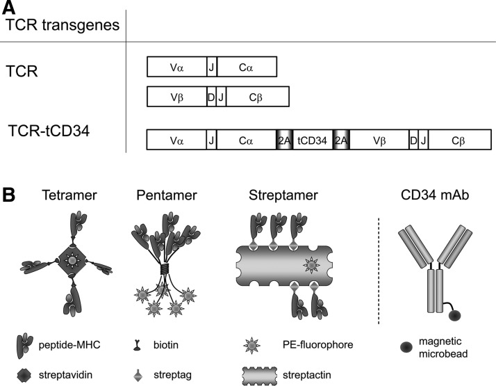
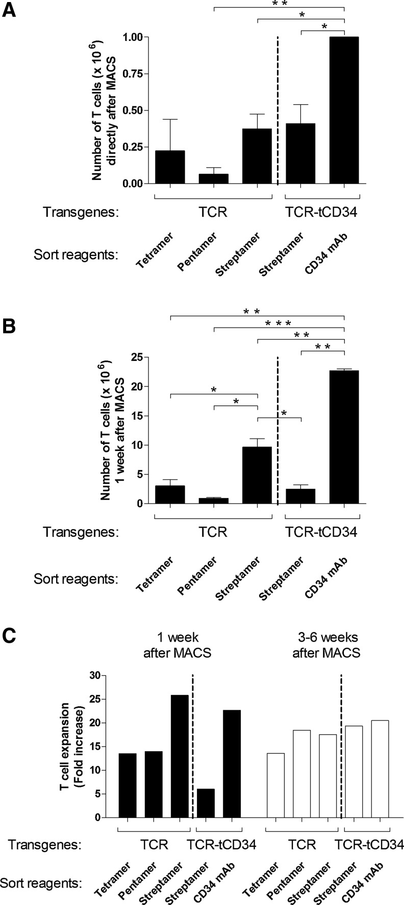
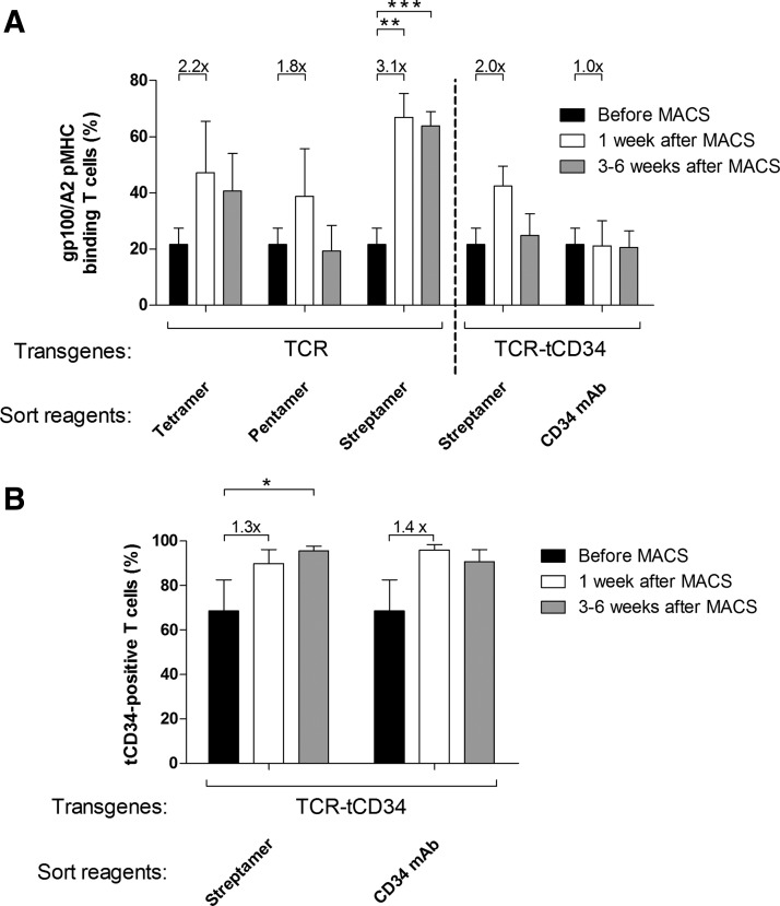
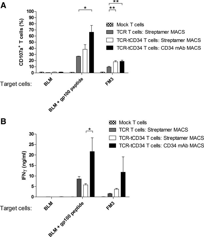
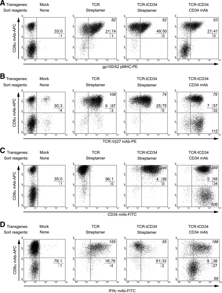
T cell-sorting technologies with peptide-MHC multimers or antibodies against gene markers enable enrichment of antigen-specific T cells and are expected to enhance the therapeutic efficacy of clinical T cell therapy. However, a direct comparison between sorting reagents for their ability to enrich T cells is lacking. Here, we compared the in vitro properties of primary human T cells gene-engineered with gp100(280-288)/HLA-A2-specific T cell receptor-αβ (TCRαβ) on magnetic-activated cell sorting (MACS) with various peptide-MHC multimers or an antibody against truncated CD34 (tCD34). With respect to peptide-MHC multimers, we observed that Streptamer(®), when compared with pentamers and tetramers, improved T cell yield as well as level and stability of enrichment, of TCR-engineered T cells (>65% of peptide-MHC-binding T cells, stable for at least 6 weeks). In agreement with these findings, Streptamer, the only detachable reagent, revealed significant T cell expansion in the first week after MACS. Sorting TCR and tCD34 gene-engineered T cells with CD34 monoclonal antibody (mAb) resulted in the most significant T cell yield and enrichment of T cells (>95% of tCD34 T cells, stable for at least 6 weeks). Notably, T cells sorted with CD34 mAb, when compared with Streptamer, bound about 2- to 3-fold less peptide-MHC but showed superior antigen-specific upregulated expression of CD107a and production of interferon (IFN)-γ. Multiparametric flow cytometry revealed that CD4(+) T cells, uniquely present in CD34 mAb-sorted T cells, contributed to enhanced IFN-γ production. Taken together, we postulate that CD34 mAb-based sorting of gene-marked T cells has benefits toward applications of T cell therapy, especially those that require CD4(+) T cells.
Figures
Similar articles
-
gp100(209-2M) peptide immunization of human lymphocyte antigen-A2+ stage I-III melanoma patients induces significant increase in antigen-specific effector and long-term memory CD8+ T cells.Clin Cancer Res. 2004 Jan 15;10(2):668-80. doi: 10.1158/1078-0432.ccr-0095-03. Clin Cancer Res. 2004. PMID: 14760090
-
Comparison of peptide-major histocompatibility complex tetramers and dextramers for the identification of antigen-specific T cells.Clin Exp Immunol. 2014 Jul;177(1):47-63. doi: 10.1111/cei.12339. Clin Exp Immunol. 2014. PMID: 24673376 Free PMC article.
-
Clonal diversity of the T-cell population responding to a dominant HLA-A2 epitope of HER-2/neu after active immunization in an ovarian cancer patient.Hum Immunol. 2002 Jul;63(7):547-57. doi: 10.1016/s0198-8859(02)00401-9. Hum Immunol. 2002. PMID: 12072190
-
MHC and T cell development.Rev Immunogenet. 1999;1(1):91-104. Rev Immunogenet. 1999. PMID: 11256575 Review.
-
More tricks with tetramers: a practical guide to staining T cells with peptide-MHC multimers.Immunology. 2015 Sep;146(1):11-22. doi: 10.1111/imm.12499. Immunology. 2015. PMID: 26076649 Free PMC article. Review.
Cited by
-
An Alternative Application of Magnetic-Activated Cell Sorting: CD45 and CD235a Based Purification of Semen and Testicular Tissue Samples.Int J Mol Sci. 2024 Mar 24;25(7):3627. doi: 10.3390/ijms25073627. Int J Mol Sci. 2024. PMID: 38612438 Free PMC article.
-
Gamma-retroviral vector design for the co-expression of artificial microRNAs and therapeutic proteins.Nucleic Acid Ther. 2014 Oct;24(5):356-63. doi: 10.1089/nat.2014.0486. Epub 2014 Jul 14. Nucleic Acid Ther. 2014. PMID: 25019196 Free PMC article.
-
An Altered gp100 Peptide Ligand with Decreased Binding by TCR and CD8α Dissects T Cell Cytotoxicity from Production of Cytokines and Activation of NFAT.Front Immunol. 2013 Sep 4;4:270. doi: 10.3389/fimmu.2013.00270. eCollection 2013. Front Immunol. 2013. PMID: 24027572 Free PMC article.
References
-
- Altman J.D. Moss P.A. Goulder P.J., et al. Phenotypic analysis of antigen-specific T lymphocytes. Science. 1996;274:94–96. - PubMed
-
- Bendle G.M. Linnemann C. Hooijkaas A.I., et al. Lethal graft-versus-host disease in mouse models of T cell receptor gene therapy. Nat. Med. 2010;16:565–570. 561p following 570. - PubMed
-
- Besser M.J. Shapira-Frommer R. Treves A.J., et al. Clinical responses in a phase II study using adoptive transfer of short-term cultured tumor infiltration lymphocytes in metastatic melanoma patients. Clin. Cancer Res. 2010;16:2646–2655. - PubMed
Publication types
MeSH terms
Substances
LinkOut - more resources
Full Text Sources
Research Materials
Miscellaneous
