. 2012 Oct;6(5):542-52.
doi: 10.1016/j.molonc.2012.06.003. Epub 2012 Jul 27.

Genotoxic stress modulates CDC25C phosphatase alternative splicing in human breast cancer cell lines

Affiliations

Genotoxic stress modulates CDC25C phosphatase alternative splicing in human breast cancer cell lines

Hélène Albert et al. Mol Oncol. 2012 Oct.

Abstract

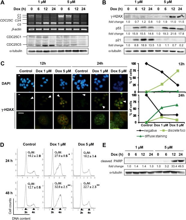
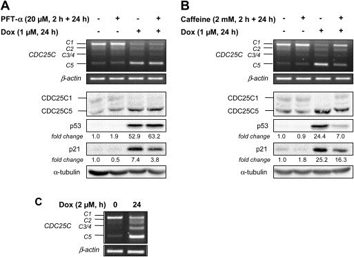
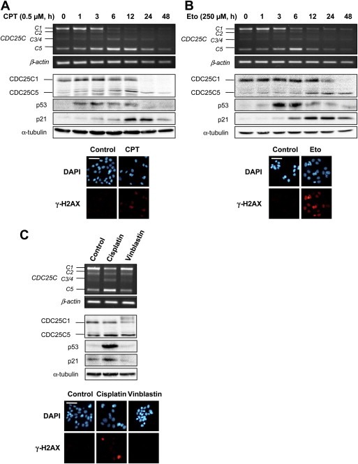
CDC25 (cell division cycle 25) phosphatases are essential for cell cycle control under normal conditions and in response to DNA damage. They are represented by three isoforms, CDC25A, B and C, each of them being submitted to an alternative splicing mechanism. Alternative splicing of many genes is affected in response to genotoxic stress, but the impact of such a stress on CDC25 splicing has never been investigated. In this study, we demonstrate that genotoxic agents (doxorubicin, camptothecin, etoposide and cisplatin), alter the balance between CDC25C splice variants in human breast cancer cell lines both at the mRNA and protein levels. This modulation occurs during the response to moderate, sub-lethal DNA damage. Our results also suggest that the CDC25C splice variants expression shift induced by a genotoxic stress is dependent on the ATM/ATR signaling but not on p53. This study highlights the modulation of CDC25C alternative splicing as an additional regulatory event involved in cellular response to DNA damage in breast cancer cells.


Figures

Figure 1
Figure 1
Modulation of CDC25C splicing by doxorubicin in human breast cancer cell lines. (A) Schematic representation of CDC25A, CDC25B and CDC25C pre‐mRNA and alternatively spliced transcripts sequences. Colored rectangles correspond to alternatively spliced exons. Arrows show the localization of primers used to amplify the different CDC25A, CDC25B and CDC25C variants by semi‐quantitative RT‐PCR (see Materials and Methods for primers sequences). Amplicon length expected for each splice variant appears at the right of the figure. (B) MCF‐7 cells were treated with 1 μM of doxorubicin at the indicated times. RNA was subjected to semi‐quantitative RT‐PCR to detect CDC25A, CDC25B and CDC25C splice variants. PCR products are identified on the left and molecular weight markers are indicated on the right. The β‐actin gene was used as a standardizing control. (C), top, RNA from MCF‐7 cells treated with doxorubicin as indicated were subjected to Real‐Time quantitative RT‐PCR to evaluate the ratio between CDC25C5 and C1 variants. The results are expressed as 2ΔCT C5/2ΔCT C1 (mean ± S.D.) of three independent experiments. 18S rRNA was used as an endogenous reference gene. ** Shows significant difference from control at p < 0.01 (Student's t test); bottom, schematic representation of primers (arrows) and TaqMan® probe (bold line) used to detect C1 and C5 variants. (D) CDC25C protein expression from MCF‐7 cells treated as indicated was examined by immunoblotting. α‐tubulin was used as a loading control. Molecular weights for each protein are indicated on the right. (E) The MCF‐7 multidrug‐resistant counterpart cell line Vcr‐R was treated with 2 μM of doxorubicin for 12 h. CDC25C splicing was studied by semi‐quantitative RT‐PCR as described above.
Figure 2
Figure 2
CDC25C splicing modulation induced by doxorubicin is associated with DNA‐damage and cell cycle arrest, independently of apoptosis. MCF‐7 cells were treated with 1 μM and 5 μM of doxorubicin for the indicated times. (A) CDC25C variants expression was evaluated by RT‐PCR (top) and immunoblotting (bottom). β‐actin and α‐tubulin were used as internal controls for the RT‐PCR and immoblotting assays, respectively. (B) Immunoblotting of DNA damage response proteins. The fold‐changes of protein levels between treatment and control, measured by densitometry, are shown beneath the blots. Equal loading was confirmed by α‐tubulin immunoblot. (C) Immunofluorescence analysis of nuclear γ‐H2AX foci formation using anti‐γ‐H2AX specific antibody (green, FITC). Images at the bottom correspond to a magnification of the cell pointed by an arrow in the images above. Nuclei were counterstained with DAPI (blue). Scale bars, 10 μm. Graphs on the right represent the count of at least 50 cells that were classified into non‐stained cells (negative), cells with discrete γ‐H2AX foci (discrete foci, at least three foci per cells) and cells with diffuse γ‐H2AX staining (diffuse staining). (D) Cell cycle distribution was monitored by flow cytometry after propidium iodide staining. Shown is the result of a representative FACS experiment and the frequency of cells in G2/M phase. Data are displayed as mean ± S.D. of three independent experiments. * and ** indicate a significant difference between treated and untreated cells with p < 0.05 and p < 0.01, respectively. (E) Analysis of PARP cleavage by immunoblotting. The fold‐changes of protein levels between treatment and control, measured by densitometry, are shown beneath the blots. Equal loading was confirmed by α‐tubulin immunoblot.
Figure 3
Figure 3
Regulation of CDC25C splicing is dependent on ATM/ATR and independent on p53 pathways. MCF‐7 cells (with wild‐type p53) were pre‐treated with 20 μM of the p53 inhibitor pifithrin‐α (PFT‐α) (A) or with 2 mM of the ATM/ATR kinases inhibitor caffeine (B) for 2 h, followed by a co‐incubation with 1 μM of doxorubicin for 24 h, top, RNA was subjected to RT‐PCR to detect CDC25C splice variants. The β‐actin gene was used as an internal control; bottom, Immunoblots were performed to evaluate CDC25C, p53 and p21 proteins expression. The fold‐changes of protein expression between treatment and control, measured by densitometry, are shown beneath the blots. Staining of the blot with α‐tubulin was used as an internal loading control. (C) MDA‐MB‐231 cells (with mutated‐p53) were treated with 2 μM of doxorubicin for 24 h and CDC25C splice variants expression was assessed by RT‐PCR. The β‐actin gene was used as an internal control.
Figure 4
Figure 4
Modulation of CDC25C alternative splicing in response to other DNA‐damaging agents in MCF‐7 cells. Cells were treated with the topoisomerase I inhibitor camptothecin (CPT, 0.5 μM) (A) and the topoisomerase II inhibitor etoposide (Eto, 250 μM) (B) for the indicated times. Cells were also treated with the chemotherapeutic agents cisplatin (50 μM) and vinblastin (1 μM) for 12 h (C). Top, RNA was subjected to semi‐quantitative RT‐PCR to detect CDC25C splice variants. The β‐actin gene was used as a standardizing control; middle, Cell lysates were subjected to immunoblotting. Equal protein loading was confirmed by α‐tubulin immunoblot; bottom, cells were treated with the different compounds for 24 h and immunostained with anti‐γ‐H2AX specific antibodies (red, AlexaFluor 594). Nuclei were counterstained with DAPI (blue). Scale bars, 50 μm.

References

    1. Albert, H. , Santos, S. , Battaglia, E. , Brito, M. , Monteiro, C. , Bagrel, D. , 2011. Differential expression of CDC25 phosphatases splice variants in human breast cancer cells. Clin. Chem. Lab. Med.. 49, 1707–1714. - PubMed
    1. Aressy, B. , Ducommun, B. , 2008. Cell cycle control by the CDC25 phosphatases. Anticancer Agents Med. Chem.. 8, 818–824. - PubMed
    1. Baldin, V. , Cans, C. , Superti-Furga, G. , Ducommun, B. , 1997. Alternative splicing of the human CDC25B tyrosine phosphatase. Possible implications for growth control?. Oncogene. 14, 2485–2495. - PubMed
    1. Bartek, J. , Iggo, R. , Gannon, J. , Lane, D.P. , 1990. Genetic and immochemical analysis of mutant p53 in human breast cancer cell lines. Oncogene. 5, 893–899. - PubMed
    1. Biamonti, G. , Caceres, J.F. , 2009. Cellular stress and RNA splicing. Trends Biochem. Sci.. 34, 146–153. - PubMed

Publication types