. 2013 Mar;19(3):233-43.
doi: 10.1089/ten.TEC.2012.0221. Epub 2012 Sep 13.

Effect of surface pore structure of nerve guide conduit on peripheral nerve regeneration

Affiliations

Effect of surface pore structure of nerve guide conduit on peripheral nerve regeneration

Se Heang Oh et al. Tissue Eng Part C Methods. 2013 Mar.

Abstract

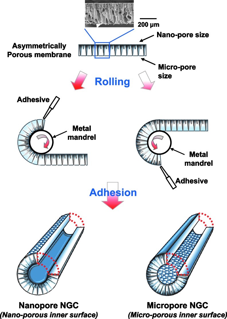
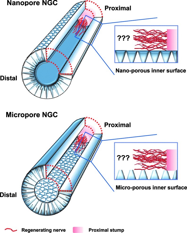
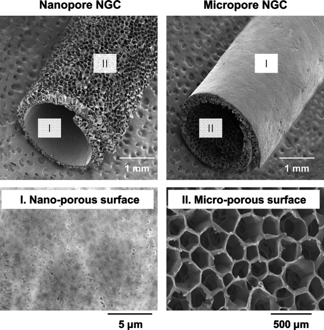
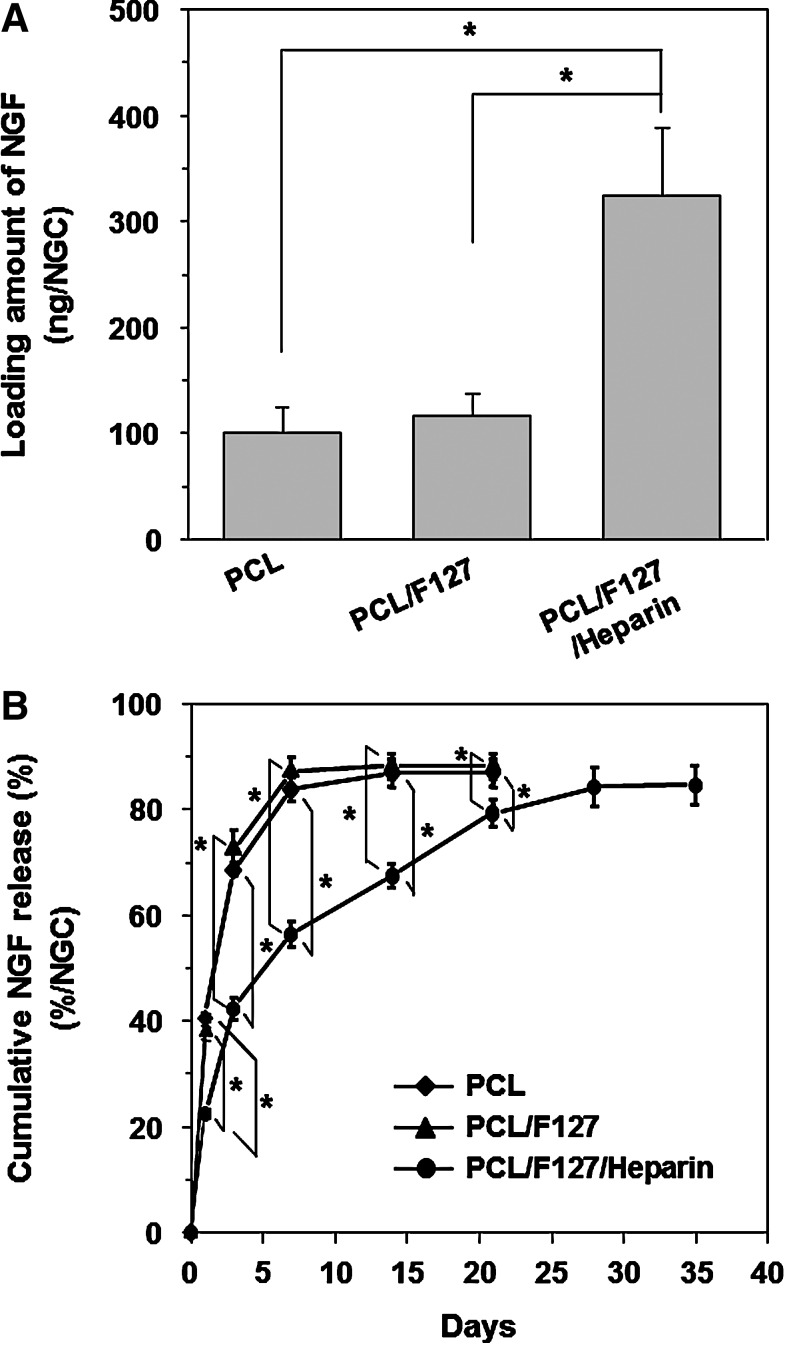
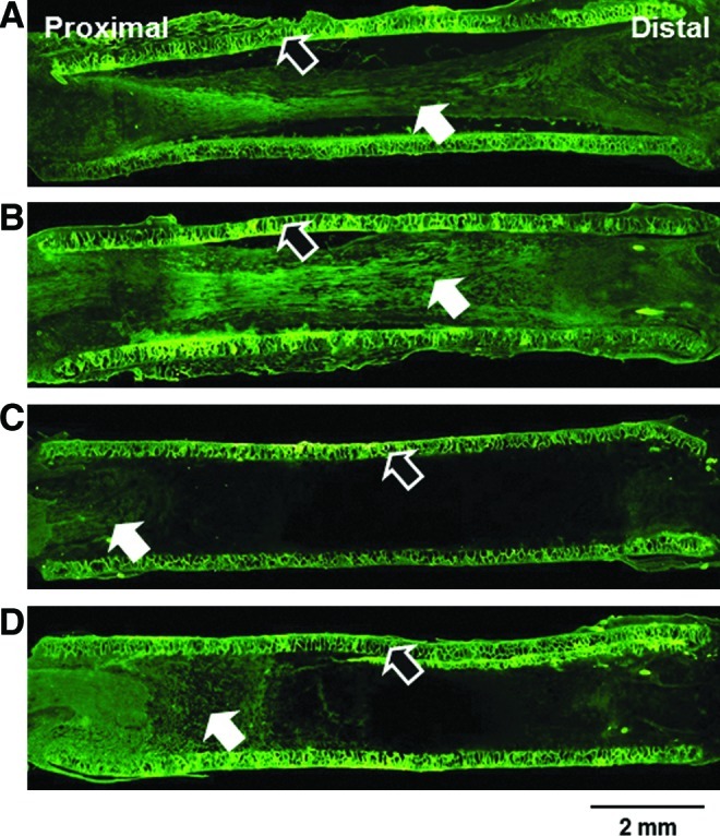
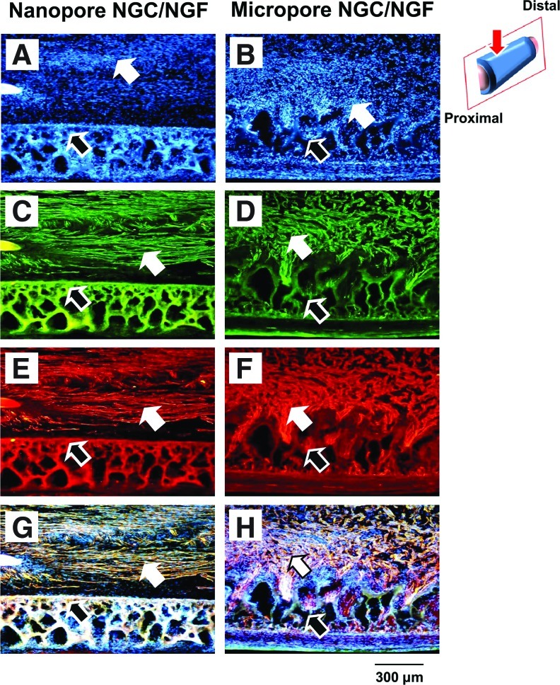
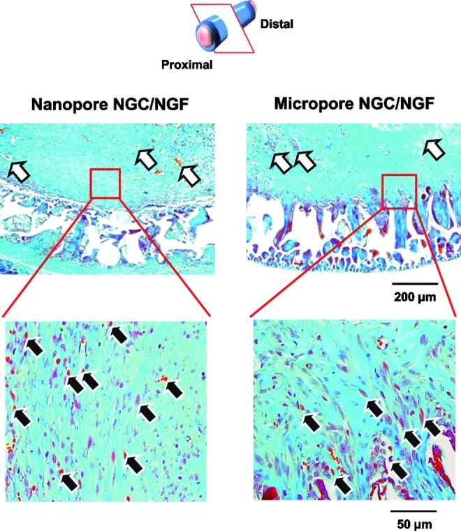
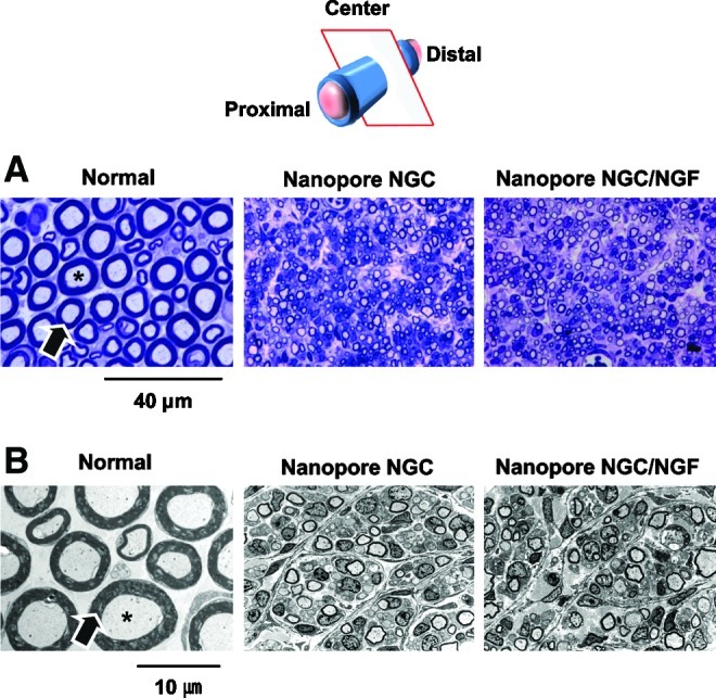
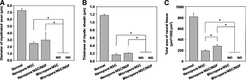
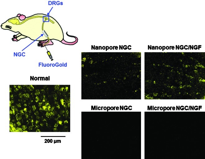
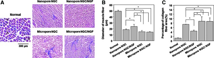
Polycaprolactone (PCL)/Pluronic F127 nerve guide conduits (NGCs) with different surface pore structures (nano-porous inner surface vs. micro-porous inner surface) but similar physical and chemical properties were fabricated by rolling the opposite side of asymmetrically porous PCL/F127 membranes. The effect of the pore structure on peripheral nerve regeneration through the NGCs was investigated using a sciatic nerve defect model of rats. The nerve fibers and tissues were shown to have regenerated along the longitudinal direction through the NGC with a nano-porous inner surface (Nanopore NGC), while they grew toward the porous wall of the NGC with a micro-porous inner surface (Micropore NGC) and, thus, their growth was restricted when compared with the Nanopore NGC, as investigated by immunohistochemical evaluations (by fluorescence microscopy with anti-neurofilament staining and Hoechst staining for growth pattern of nerve fibers), histological evaluations (by light microscopy with Meyer's modified trichrome staining and Toluidine blue staining and transmission electron microscopy for the regeneration of axon and myelin sheath), and FluoroGold retrograde tracing (for reconnection between proximal and distal stumps). The effect of nerve growth factor (NGF) immobilized on the pore surfaces of the NGCs on nerve regeneration was not so significant when compared with NGCs not containing immobilized NGF. The NGC system with different surface pore structures but the same chemical/physical properties seems to be a good tool that is used for elucidating the surface pore effect of NGCs on nerve regeneration.


Figures

FIG. 1.
FIG. 1.
Schematic diagrams showing the process used to fabricate the NGCs with different surface pore structures (nano-porous inner surface vs. micro-porous inner surface) by rolling the opposite side of the asymmetrically porous PCL/Pluronic F127 membranes. NGC, nerve guide conduits; PCL, polycaprolactone. Color images available online at www.liebertpub.com/tec
FIG. 2.
FIG. 2.
Schematic diagrams showing the Nanopore NGC (with nano-porous inner surface) and Micropore NGC (with micro-porous inner surface), and their nerve regeneration potential through the NGCs. Color images available online at www.liebertpub.com/tec
FIG. 3.
FIG. 3.
Scanning electron microscope photographs showing the gross appearance and surface morphology of the Nanopore NGC and Micropore NGC prepared by rolling the opposite side of the asymmetrically porous PCL/F127 (5 wt.%) membranes.
FIG. 4.
FIG. 4.
(A) Loading and (B) cumulative released amounts of NGF from the PCL, PCL/F127, and PCL/F127/heparin NGCs (n=3; *p<0.05). The heparin-immobilized NGC (PCL/F127/heparin) had a significantly higher loading amount of NGF than the NGCs without immobilized heparin (PCL and PCL/F127), which was probably due to the ionic interactions between heparin and NGF molecules. The PCL/F127/heparin NGC showed a moderate burst release of NGF at the initial stage (for 3 days), and then, the NGF was continuously released over 35 days. NGF, nerve growth factor.
FIG. 5.
FIG. 5.
Longitudinal sections of the regenerated nerve through (A) Nanopore NGC, (B) Nanopore NGC/NGF, (C) Micropore NGC, and (D) Micropore NGC/NGF (4 weeks after implantation; anti-neurofilament staining [NF-200],×4; white arrow, regenerated nerve; black arrow, NGC). The Nanopore NGCs showed faster axonal growth than the Micropore NGCs, regardless of the presence of NGF. Color images available online at www.liebertpub.com/tec
FIG. 6.
FIG. 6.
Longitudinal sections of the regenerated nerve fibers through the Nanopore NGC/NGF and Micropore NGC/NGF. (A, B) Hoechst staining; (C, D) NF-200 staining; (E, F) S100β staining; and (G, H) their merged images (4 weeks after implantation;×100; white arrow, regenerated nerve fiber; black arrow, NGC; red arrow, observation position). For the Nanopore NGC/NGF, the migrating cells were predominantly positioned at the central region in the NGC without attachment on the conduit wall surface. The regenerating nerve fibers were arranged along the longitudinal direction of the conduit. In contrast, for the Micropore NGC/NGF, the cells were scattered without any arrangement, and the regenerated nerve fibers had infiltrated into the porous wall of the conduit. Color images available online at www.liebertpub.com/tec
FIG. 7.
FIG. 7.
Transverse sections of the regenerated nerve fibers in the NGCs (4 weeks after implantation; 3 mm position from the proximal end; Meyer's trichrome staining;×100 and×400; white arrow, blood vessel; black arrow, regenerated axon). For the Nanopore NGC/NGF, axon fiber spots created by the perpendicular section of axonal fibers (red color) were observed in the regenerated nerve tissues, indicating that axonal sprouting toward the distal stump occurred. However, for the Micropore NGC/NGF, oblique-shaped axon fibers created by the slanting section of axonal fibers were detected (many of them, inside the porous wall of the conduit), suggesting that the axonal sprouting toward the porous wall of the conduit occurred. Color images available online at www.liebertpub.com/tec
FIG. 8.
FIG. 8.
(A) Light micrographs of semi-thin sections (Toluidine blue staining;×1000) and (B) Transmission electron microscope images of ultrathin sections (×3000) showing myelinated axons at the mid-portion of the Nanopore NGC and NGC/NGF 4 weeks after implantation (asterisk, axon; arrow, myelin). The Nanopore NGCs allowed for the nerve regeneration within the conduit. The Nanopore NGC/NGF showed a slightly (but no significantly) larger axon diameter and a thicker myelin sheath than the Nanopore NGC without NGF. Color images available online at www.liebertpub.com/tec
FIG. 9.
FIG. 9.
Comparison of (A) the diameter of the myelinated axon, (B) thickness of myelin sheath, and (C) area occupied by neural tissue in the NGC at the middle section 4 weeks after implantation (n=3; *p<0.05; N/D, no detection). The Nanopore NGCs show significantly faster nerve regeneration compared with the Micropore NGCs.
FIG. 10.
FIG. 10.
Fluorescent micrographs following FluoroGold (FG) retrograde tracing in the DRGs of the NGC-implanted group 1 week after FG injection (×100). The Nanopore NGC/NGF group contained more FG-labeled neuron cells compared with the Nanopore NGC group, indicating that a greater number of nerve fibers connecting the defect stumps were present. However, FG-labeled cells in the DRGs were not detected in the Micropore NGC groups, even for the NGF-immobilized NGC group. DRG, dorsal root ganglions. Color images available online at www.liebertpub.com/tec
FIG. 11.
FIG. 11.
(A) Light micrographs of gastrocnemius muscles (Masson's trichrome staining;×100), and their (B) diameter of muscle fibers and (C) percentage of collagen fiber area (n=3; *p<0.05). The Nanopore NGCs showed significantly faster recovery from muscle atrophy (thicker diameter of muscle fiber and smaller collagen fiber area) caused by the reinnervation of muscle than the Micropore NGCs. Color images available online at www.liebertpub.com/tec

Similar articles

Cited by

References

    1. Zhang N. Yan H. Wen X. Tissue-engineering approaches for axonal guidance. Brain Res Brain Res Rev. 2005;49:48. - PubMed
    1. Seckel B.R. Enhancement of peripheral nerve regeneration. Muscle Nerve. 1990;13:785. - PubMed
    1. Schlosshauer B. Müller E. Schröder B. Planck H. Müller H.W. Rat Schwann cells in bioresorbable nerve guides to promote and accelerate axonal regeneration. Brain Res. 2003;963:321. - PubMed
    1. Glück T. Ueber neuroplastik aut dem wege der transplantation. Arch Klin Chir. 1880;25:606.
    1. Maquet V. Martin D. Malgrange B. Franzen R. Schoenen J. Moonen G., et al. Peripheral nerve regeneration using bioresorbable macroporous polylactide scaffolds. J Biomed Mater Res. 2000;52:639. - PubMed

Publication types

MeSH terms