. 2012 Aug 7;5(236):ra56.
doi: 10.1126/scisignal.2002829.

Microtubules underlie dysfunction in duchenne muscular dystrophy

Affiliations

Microtubules underlie dysfunction in duchenne muscular dystrophy

Ramzi J Khairallah et al. Sci Signal. .

Abstract

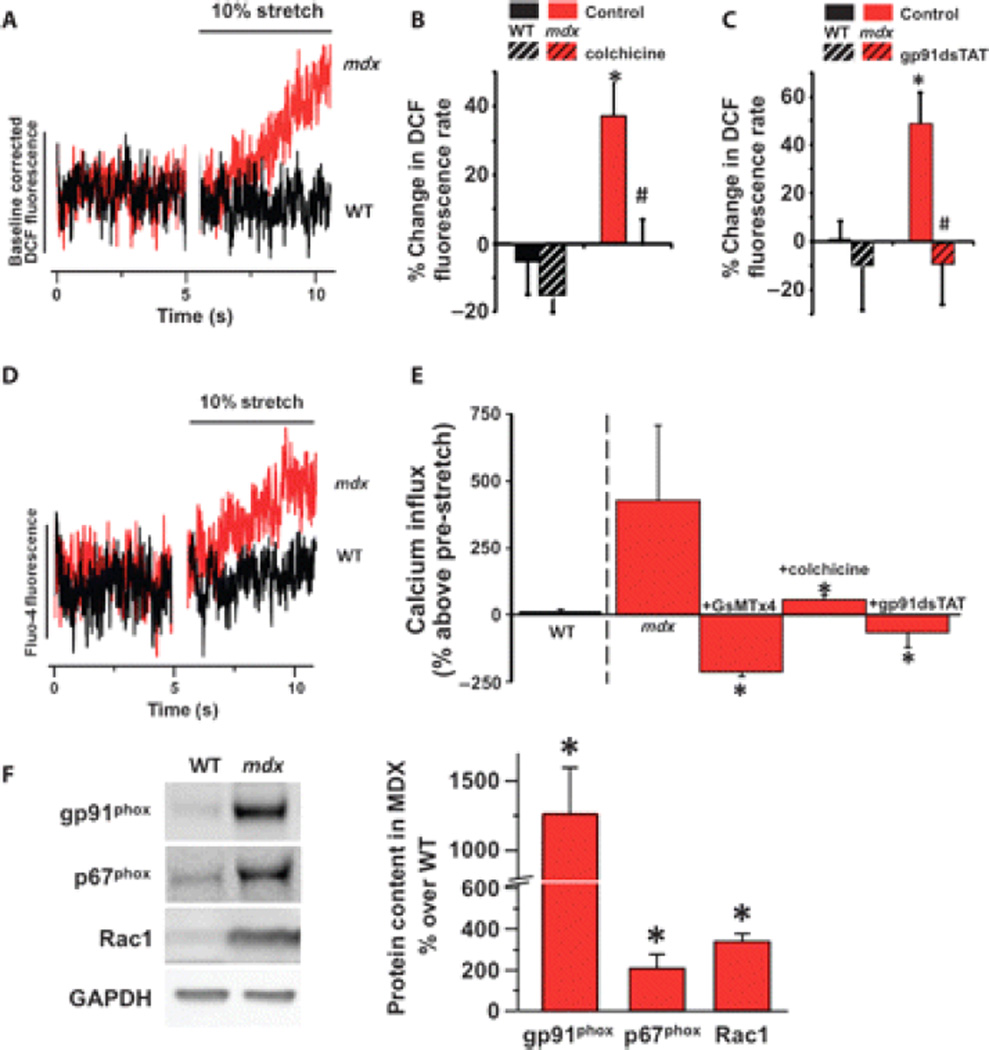
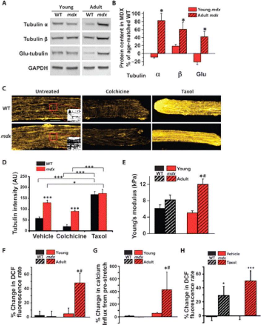
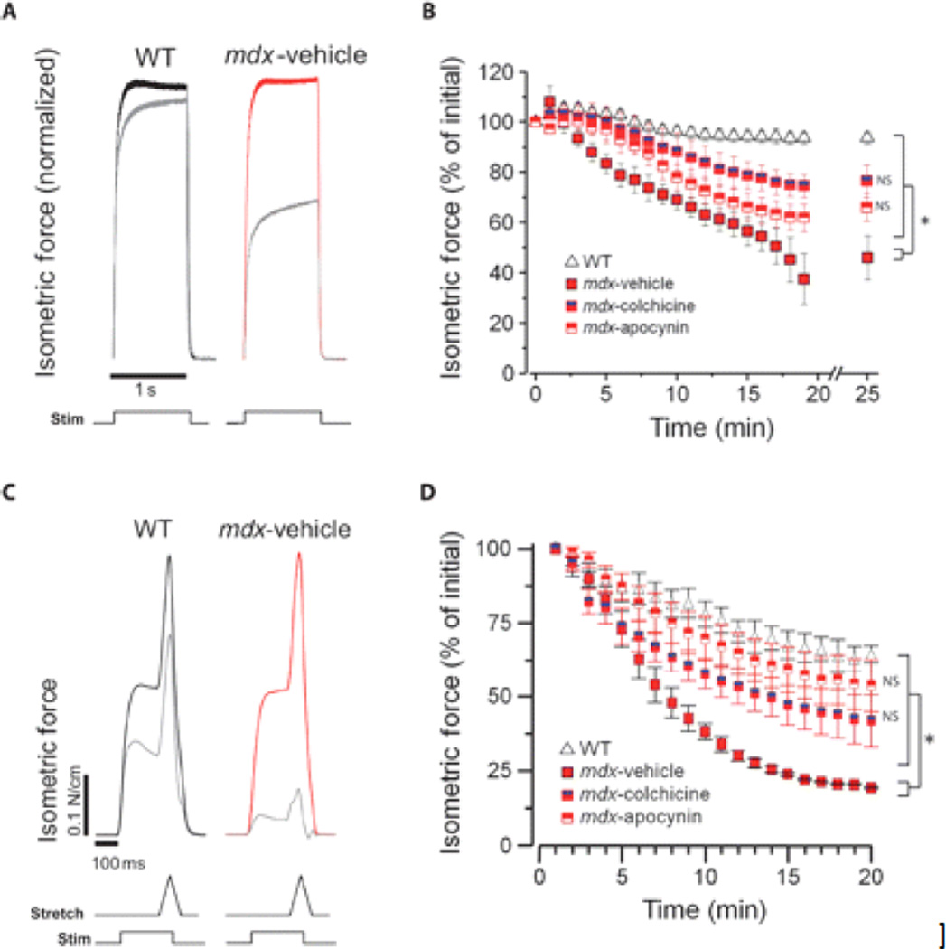
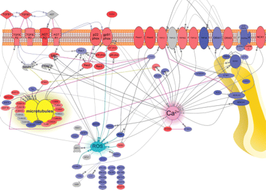
Duchenne muscular dystrophy (DMD) is a fatal X-linked degenerative muscle disease caused by the absence of the microtubule-associated protein dystrophin, which results in a disorganized and denser microtubule cytoskeleton. In addition, mechanotransduction-dependent activation of calcium (Ca(2+)) and reactive oxygen species (ROS) signaling underpins muscle degeneration in DMD. We show that in muscle from adult mdx mice, a model of DMD, a brief physiologic stretch elicited microtubule-dependent activation of NADPH (reduced-form nicotinamide adenine dinucleotide phosphate) oxidase-dependent production of ROS, termed X-ROS. Further, X-ROS amplified Ca(2+) influx through stretch-activated channels in mdx muscle. Consistent with the importance of the microtubules to the dysfunction in mdx muscle, muscle cells with dense microtubule structure, such as those from adult mdx mice or from young wild-type mice treated with Taxol, showed increased X-ROS production and Ca(2+) influx, whereas cells with a less dense microtubule network, such as young mdx or adult mdx muscle treated with colchicine or nocodazole, showed little ROS production or Ca(2+) influx. In vivo treatments that disrupted the microtubule network or inhibited NADPH oxidase 2 reduced contraction-induced injury in adult mdx mice. Furthermore, transcriptome analysis identified increased expression of X-ROS-related genes in human DMD skeletal muscle. Together, these data show that microtubules are the proximate element responsible for the dysfunction in Ca(2+) and ROS signaling in DMD and could be effective therapeutic targets for intervention.


Figures

Fig. 1
Fig. 1
X-ROS signaling in dystrophic muscle fibers. (A) Representative averaged data of adult (5 to 6 months) wild-type (WT; n = 5) and mdx (n = 5) single FDB myofibers loaded with DCF and subjected to a 10% stretch. The DCF fluorescence profiles pre- and post-stretch were fit by linear least squares regression. The rates of DCF fluorescence pre- and post-stretch were taken as the rate of ROS production. The DCF fluorescence change was normalized to the pre-stretch to allow comparison of the post-stretch rates. (B) The microtubule polymerization inhibitor colchicine also prevented the increase in X-ROS in mdx. (C) NOX2 inhibition abrogates stretch-induced ROS production in mdx fibers. Fibers from WT (n = 3 animals, 32 fibers) and mdx (n = 3 animals, 23 fibers) were subjected to acute stretch with a scrambled peptide or with the NOX2 inhibitor gp91dsTAT. Mean values of DCF fluorescence, calculated as percent change over pre-stretch rate, were significantly increased in mdx (*P < 0.05) compared to WT. In mdx fibers, stretch ROS production was inhibited by gp91dsTAT (#P < 0.05). (D) Representative averaged data of 5- to 6-month adult WT and mdx single FDB myofibers loaded with fluo-4. (E) Acute membrane stretch increases sarcolemmal calcium influx in mdx myofibers, as measured by fluo-4 fluorescence rate increase, and is prevented by the SAC inhibitor GsMTx4 (n = 2 animals, 10 fibers), colchicine (n = 3 animals, 12 fibers), and gp91dsTAT (n = 2 animals, 8 fibers) (*P < 0.05 compared to mdx). The increase in the myoplasmic concentration of Ca2+ with stretch was independent of sarcoplasmic reticulum release because inhibition of ryanodine receptors did not block Ca2+ influx (−0.005 ± 0.046 pre-stretch rate compared to 0.751 ± 0.296 stretch rate; P < 0.01 paired t test). (F) Western blot analysis of mdx tibialis anterior muscle displays increased abundance of the NOX2 subunits gp91phox, p67phox, and Rac1 (t test; *P < 0.05 compared to WT).
Fig. 2
Fig. 2
Only adult dystrophic muscle displays increased density of the microtubule network, an upstream modulator of X-ROS. (A and B) Adult, but not young, mdx muscle shows increased abundance of the tubulin subunits α and β and increased detyrosination of tubulin (Glu-tubulin) compared to age-matched WT muscle (*P < 0.05 compared to young; n = 6 animals per genotype per age). (C) Immunohistochemistry of α-tubulin reveals that mdx FDB shows a denser microtubule network than WT FDB. Inset panels show binarization of the region of interest (boxed in red). Scale bar, 20 µm. (D) Quantification was performed on binary images of α-tubulin immunohistochemistry (fig. S5; n = 2 animals, 12 fibers per genotype and condition; *P < 0.05, ***P < 0.001). (E) Increased microtubule network density is accompanied by increased membrane stiffness as measured by AFM (*P < 0.05 compared to young; #P < 0.05 compared to WT). (F and G) Only adult mdx muscle displays (F) X-ROS (n = 2 animals, 14 fibers per genotype; *P < 0.05 compared to young; #P < 0.05 compared to WT) and (G) increased calcium influx (n = 2 animals, 10 fibers per genotype; *P < 0.05 compared to young; #P < 0.05 compared to WT). Data from adult WT and mdx were replotted from Fig. 1, B and D, for comparison. (H) Increasing the microtubule density of young WT and mdx FDBs reveals X-ROS–competent fibers (n = 2 animals, 11 to 14 fibers per genotype; *P < 0.05, ***P < 0.001).
Fig. 3
Fig. 3
In vivo inhibition of X-ROS decreases contraction-induced injury in mdx. (A to D) EDL muscle from mdx mice treated in vivo with vehicle, colchicine, or apocynin was assayed for susceptibility to either in vitro isometric contraction–induced injury (A and B) or in vivo eccentric contraction–induced injury (C and D). Normalized isometric force transients of the initial (t = 0; WT in black, mdx in red) and final (gray) contractions (A and C). WT muscle (n = 4) exhibited little force loss after this protocol, whereas mdx (n = 3) exhibited significant force deficits. Treatment with colchicine (n = 4) or apocynin (n = 4) resulted in a significant protection from force loss (ANOVA: *P < 0.05; NS, not significant compared to WT) (B). WT muscle (n = 3) displayed an ~40% decrease in specific force generation after 20 eccentric contractions, whereas force generation was almost absent in mdx muscle. Treatment with colchicine (n = 4) or apocynin (n = 3) resulted in a significant protection from force loss (ANOVA: *P < 0.05; NS, not significant compared to WT) (D).
Fig. 4
Fig. 4
Transcriptome analysis of DMD muscle supports the X-ROS signaling mechanism in DMD. Genes in red show significantly increased expression and those in blue show significantly decreased expression in DMD muscle. Arrows represent direct positive regulation. Blunt arrows represent negative regulation. AGT, angiotensin II; AGTR, angiotensin II receptor; CALM, calmodulin; CASQ, calsequestrin; CAT, catalase; CAV3, caveolin 3; CUL3, cullin 3; DHPR, dihydropyridine receptor; GLRX, glutaredoxin; GSR, glutathione S-reductase; GST, glutathione S-transferase; JCTN, junction; MAO, monoamine oxidase; MAP, microtubule-associated protein; NOS, nitric oxide synthase; PLN, phospholamban; PRDX, peroxiredoxin; PRKCA, protein kinase Cα; SERCA, sarcoplasmic/endoplasmic reticulum calcium ATPase; SOD, superoxide dismutase; STMN1, stathmin 1; TGFBR, transforming growth factor–β receptor; TRDN, triadin; TUBA, tubulin α; TUBB, tubulin β; TXN, thioredoxin; TXNRD, thioredoxin reductase. These results from clinical patient samples suggest that the X-ROS signaling pathway operates in human DMD muscle.

References

    1. Hoffman EP, Brown RH, Jr, Kunkel LM. Dystrophin: The protein product of the Duchenne muscular dystrophy locus. Cell. 1987;51:919–928. - PubMed
    1. Pegoraro E, Hoffman EP, Piva L, Gavassini BF, Cagnin S, Ermani M, Bello L, Soraru G, Pacchioni B, Bonifati MD, Lanfranchi G, Angelini C, Kesari A, Lee I, Gordish-Dressman H, Devaney JM, McDonald CM. SPP1 genotype is a determinant of disease severity in Duchenne muscular dystrophy. Neurology. 2011;76:219–226. - PMC - PubMed
    1. Nishiyama A, Takeshima Y, Saiki K, Narukage A, Oyazato Y, Yagi M, Matsuo M. Two novel missense mutations in the myostatin gene identified in Japanese patients with Duchenne muscular dystrophy. BMC Med. Genet. 2007;8:19. - PMC - PubMed
    1. Allen DG, Gervasio OL, Yeung EW, Whitehead NP. Calcium and the damage pathways in muscular dystrophy. Can. J. Physiol. Pharmacol. 2010;88:83–91. - PubMed
    1. Whitehead NP, Yeung EW, Froehner SC, Allen DG. Skeletal muscle NADPH oxidase is increased and triggers stretch-induced damage in the mdx mouse. PLoS One. 2010;5:e15354. - PMC - PubMed

Publication types

MeSH terms

LinkOut - more resources