

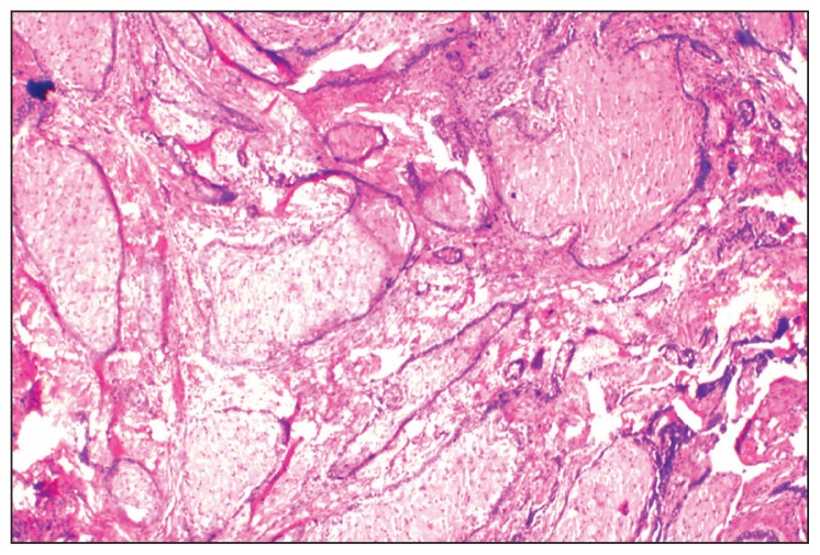
Granular cell ameloblastoma showing desmoplasia
- PMID: 22871627
- PMCID: PMC6081002
- DOI: 10.5144/0256-4947.2012.30.5.1342
Granular cell ameloblastoma showing desmoplasia
Abstract
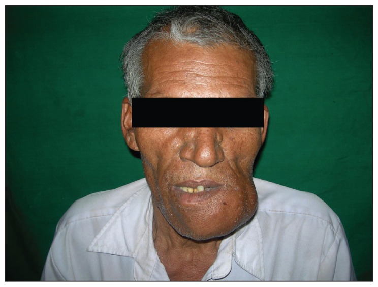
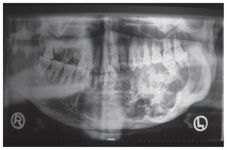
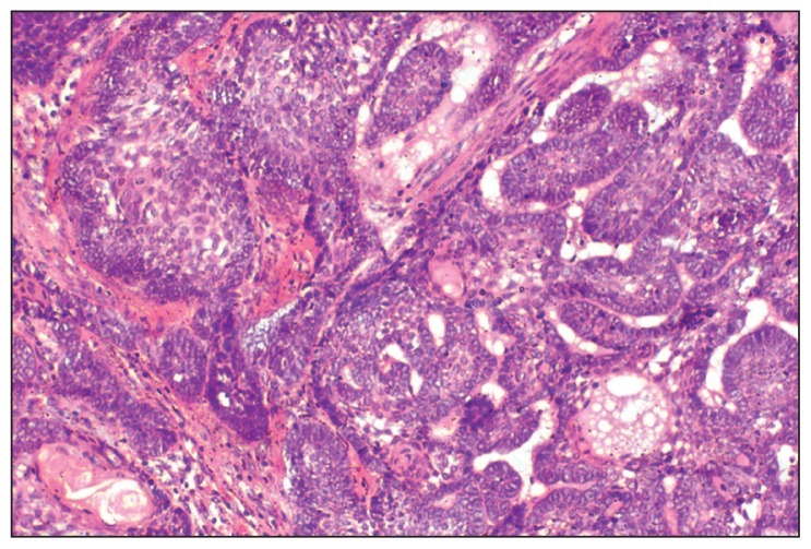
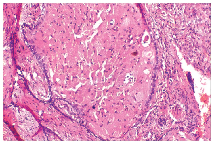
Our case of ameloblastoma had a surprisingly long 25 year history, with abnormally large dimensions, a multilocular diffuse-mixed radiographic picture, and was histopathologically diagnosed as granular cell ameloblastoma with desmoplasia. To the best of our knowledge, this is the first ameloblastoma ever reported, that has shown combined features of granular cells, desmoplasia, ameloblastic follicles, plexiform, and acanthomatous patterns. The nature of granular cells in this type of tumor and the significance of their presence have also been reviewed. From the studies on ameloblastomas to date, it seems that the old belief that granular cell ameloblastoma is the most aggressive variant of ameloblastoma is a myth, and in all probability, granular cells are just a transitional or matured phase in the life cycle of ameloblastomas, starting with normal stellate reticulum-like cells, leading to a production of granules and finally leading to degeneration and formation of cystic areas.
Figures
References
-
- Cranin AN, Benett J, Solomon M, Quarcoo S. Massive Granular cell ameloblastoma with metastasis: Report of a case. J Oral Maxillofac Surg. 1987;45:800–4. - PubMed
-
- Nasu M, Tagaki M, Yamamoto H. Ultrastructural and histochemical studies of granular cell ameloblastoma. J Oral Pathol. 1984;13:448–56. - PubMed
-
- Tandler B, Rossi EP. Granular cell ameloblastoma: Electron microscopic observations. J Oral Pathol. 1977;6:401–12. - PubMed
-
- Kumamoto H, Kimi K, Ooya K. Immunohistochemical and ultrastructural investigation of apoptotic cell death in Granular cell ameloblastoma. J Oral Pathol Med. 2001;30:245–50. - PubMed
-
- Ara SG, Han PP, Tamamura R, Nagatsuka H, Hu H, Katase N, et al. Immunolocalization of cell signaling molecules in the granular cell ameloblastoma. J Oral Pathol Med. 2007;36:609–14. - PubMed
Publication types
MeSH terms
LinkOut - more resources
Full Text Sources