

Influence of select extracellular matrix proteins on mesenchymal stem cell osteogenic commitment in three-dimensional contexts
- PMID: 22871641
- PMCID: PMC3488129
- DOI: 10.1016/j.actbio.2012.07.048
Influence of select extracellular matrix proteins on mesenchymal stem cell osteogenic commitment in three-dimensional contexts
Abstract
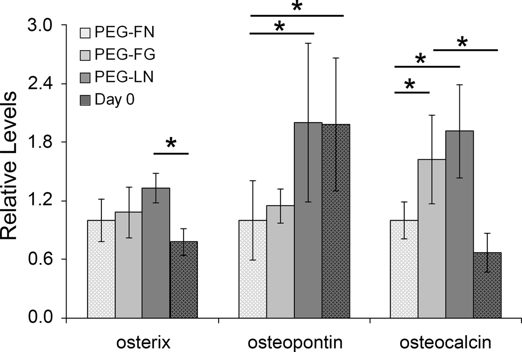
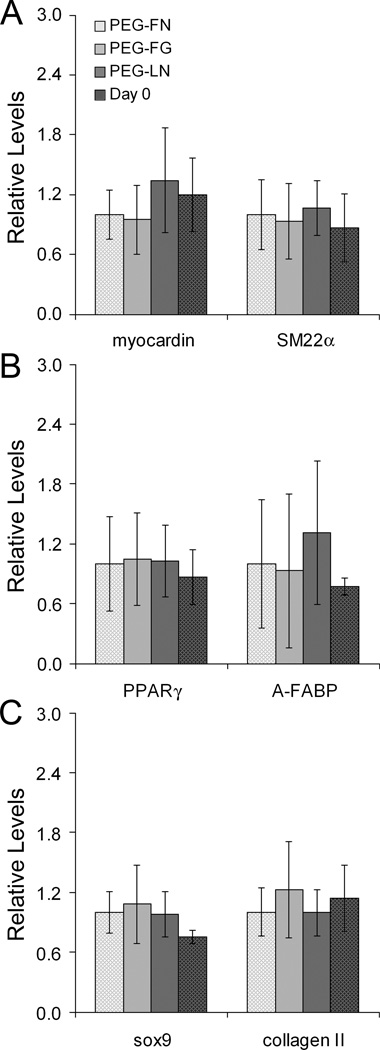
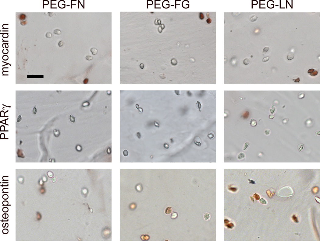
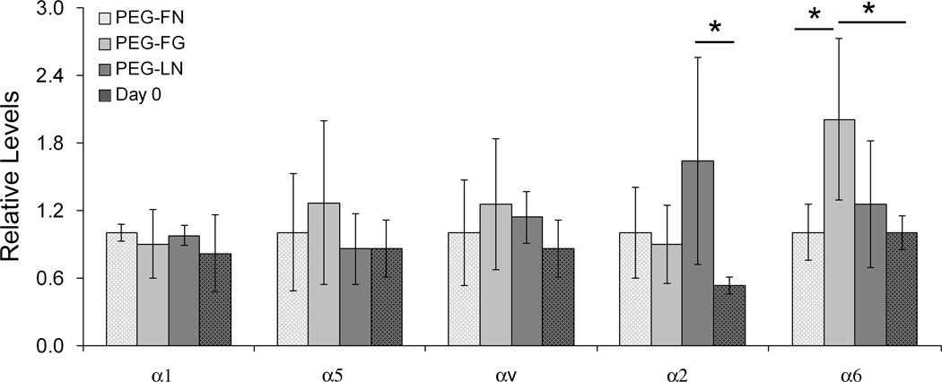
Growth factors have been shown to be powerful mediators of mesenchymal stem cell (MSC) osteogenic differentiation. However, their use in tissue engineered scaffolds not only can be costly but also can induce undesired responses in surrounding tissues. Thus, the ability to specifically promote MSC osteogenic differentiation in the absence of exogenous growth factors via the manipulation of scaffold material properties would be beneficial. The current work examines the influence of select extracellular matrix (ECM) proteins on MSC osteogenesis toward the goal of developing scaffolds with intrinsically osteoinductive properties. Fibrinogen (FG), fibronectin (FN) and laminin-1 (LN) were chosen for evaluation due to their known roles in bone morphogenesis or bone fracture healing. These proteins were conjugated into poly(ethylene glycol) diacrylate (PEGDA) hydrogels and their effects on encapsulated 10T½ MSCs were evaluated. Specifically, following 1week of culture, mid-term markers of various MSC lineages were examined in order to assess the strength and specificity of the observed osteogenic responses. PEG-LN gels demonstrated increased levels of the osteogenic transcription factor osterix relative to day 0 levels. In addition, PEG-FG and PEG-LN gels were associated with increased deposition of bone ECM protein osteocalcin relative to PEG-FN gels and day 0. Importantly, the osteogenic response associated with FG and LN appeared to be specific in that markers for chondrocytic, smooth muscle cell and adipocytic lineages were not similarly elevated relative to day 0 in these gels. To gain insight into the integrin dynamics underlying the observed differentiation results, initial integrin adhesion and temporal alterations in cell integrin profiles were evaluated. The associated results suggest that α(2), α(v) and α(6) integrin subunits may play key roles in integrin-mediated osteogenesis.
Copyright © 2012 Acta Materialia Inc. Published by Elsevier Ltd. All rights reserved.
Figures
References
-
- Kundu AK, Putnam AJ. Vitronectin and collagen I differentially regulate osteogenesis in mesenchymal stem cells. Biochemical and Biophysical Research Communications. 2006;347:347–357. - PubMed
Publication types
MeSH terms
Substances
Grants and funding
LinkOut - more resources
Full Text Sources
Molecular Biology Databases
Miscellaneous
