

The MEN mediates the effects of the spindle assembly checkpoint on Kar9-dependent spindle pole body inheritance in budding yeast
- PMID: 22871738
- PMCID: PMC3442921
- DOI: 10.4161/cc.21504
The MEN mediates the effects of the spindle assembly checkpoint on Kar9-dependent spindle pole body inheritance in budding yeast
Abstract
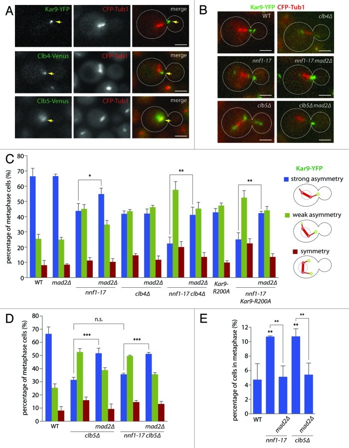
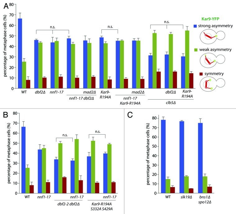
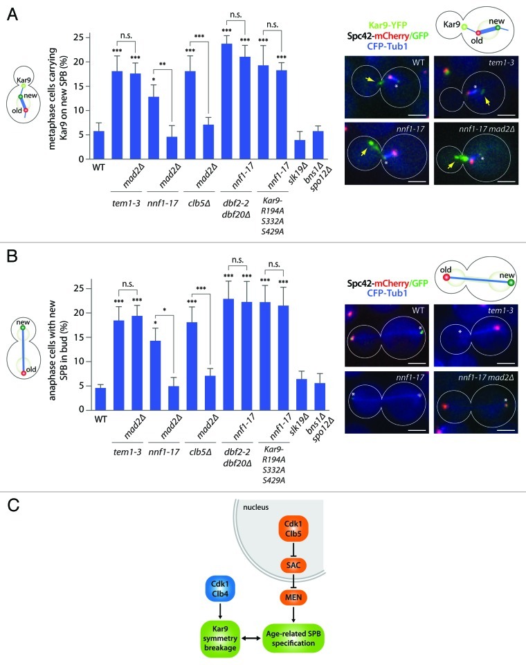
Many asymmetrically dividing cells segregate the poles of the mitotic spindle non-randomly between their two daughters. In budding yeast, the protein Kar9 localizes almost exclusively to the astral microtubules emanating from the old spindle pole body (SPB) and promotes its movement toward the bud. Thereby, Kar9 orients the spindle relative to the division axis. Here, we show that beyond perturbing Kar9 distribution, activation of the spindle assembly checkpoint (SAC) randomizes SPB inheritance. Inactivation of the B-type cyclin Clb5 led to a SAC-dependent defect in Kar9 orientation and SPB segregation. Furthermore, unlike the Clb4-dependent pathway, the Clb5- and SAC-dependent pathways functioned genetically upstream of the mitotic exit network (MEN) in SPB specification and Kar9-dependent SPB inheritance. Together, our study indicates that Clb5 functions in spindle assembly and that the SAC controls the specification and inheritance of yeast SPBs through inhibition of the MEN.
Figures
Similar articles
-
Spindle pole bodies exploit the mitotic exit network in metaphase to drive their age-dependent segregation.Cell. 2012 Mar 2;148(5):958-72. doi: 10.1016/j.cell.2012.01.041. Cell. 2012. PMID: 22385961 Free PMC article.
-
Asymmetric loading of Kar9 onto spindle poles and microtubules ensures proper spindle alignment.Cell. 2003 Feb 21;112(4):561-74. doi: 10.1016/s0092-8674(03)00119-3. Cell. 2003. PMID: 12600318
-
Kar9 asymmetrical loading on spindle poles mediates proper spindle alignment in budding yeast.Dev Cell. 2003 Mar;4(3):289-90. doi: 10.1016/s1534-5807(03)00065-0. Dev Cell. 2003. PMID: 12636909 Review.
-
Activation of the DNA damage checkpoint perturbs asymmetric localization of Kar9 to spindle pole bodies in Saccharomyces cerevisiae.Biochem Biophys Res Commun. 2023 Dec 10;685:149157. doi: 10.1016/j.bbrc.2023.149157. Epub 2023 Oct 26. Biochem Biophys Res Commun. 2023. PMID: 37918324
-
Coupling spindle position with mitotic exit in budding yeast: The multifaceted role of the small GTPase Tem1.Small GTPases. 2015 Oct 2;6(4):196-201. doi: 10.1080/21541248.2015.1109023. Small GTPases. 2015. PMID: 26507466 Free PMC article. Review.
Cited by
-
Characterization of a novel separase-interacting protein and candidate new securin, Eip1p, in the fungal pathogen Candida albicans.Mol Biol Cell. 2019 Sep 1;30(19):2469-2489. doi: 10.1091/mbc.E18-11-0696. Epub 2019 Aug 14. Mol Biol Cell. 2019. PMID: 31411946 Free PMC article.
-
Characterization of a novel interaction of the Nup159 nucleoporin with asymmetrically localized spindle pole body proteins and its link with autophagy.PLoS Biol. 2023 Aug 3;21(8):e3002224. doi: 10.1371/journal.pbio.3002224. eCollection 2023 Aug. PLoS Biol. 2023. PMID: 37535687 Free PMC article.
-
Moonlighting at the Poles: Non-Canonical Functions of Centrosomes.Front Cell Dev Biol. 2022 Jul 14;10:930355. doi: 10.3389/fcell.2022.930355. eCollection 2022. Front Cell Dev Biol. 2022. PMID: 35912107 Free PMC article. Review.
-
A Novel Hyperactive Nud1 Mitotic Exit Network Scaffold Causes Spindle Position Checkpoint Bypass in Budding Yeast.Cells. 2021 Dec 24;11(1):46. doi: 10.3390/cells11010046. Cells. 2021. PMID: 35011608 Free PMC article.
-
Unifying the mechanism of mitotic exit control in a spatiotemporal logical model.PLoS Biol. 2020 Nov 12;18(11):e3000917. doi: 10.1371/journal.pbio.3000917. eCollection 2020 Nov. PLoS Biol. 2020. PMID: 33180788 Free PMC article.
References
Publication types
MeSH terms
Substances
LinkOut - more resources
Full Text Sources
Molecular Biology Databases