

Histopathological changes in the human posterior cruciate ligament during aging and osteoarthritis: correlations with anterior cruciate ligament and cartilage changes
- PMID: 22872023
- PMCID: PMC3538921
- DOI: 10.1136/annrheumdis-2012-201730
Histopathological changes in the human posterior cruciate ligament during aging and osteoarthritis: correlations with anterior cruciate ligament and cartilage changes
Abstract
Objectives: To determine the histological patterns of posterior cruciate ligament (PCL) degeneration during aging and in relation to changes in articular cartilage and anterior cruciate ligament (ACL) across the entire adult age spectrum.
Methods: Human knee joints (n=120 from 65 donors) were processed within 72 h of postmortem. Articular cartilage surfaces were graded macroscopically. Each PCL was histologically evaluated for inflammation, mucinous changes, chondroid metaplasia, cystic changes and orientation of collagen fibres. The severity of PCL degeneration was classified as normal, mild, moderate or severe. PCL scores were compared to ACL and cartilage scores from the same knees.
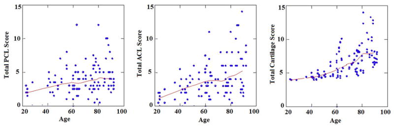
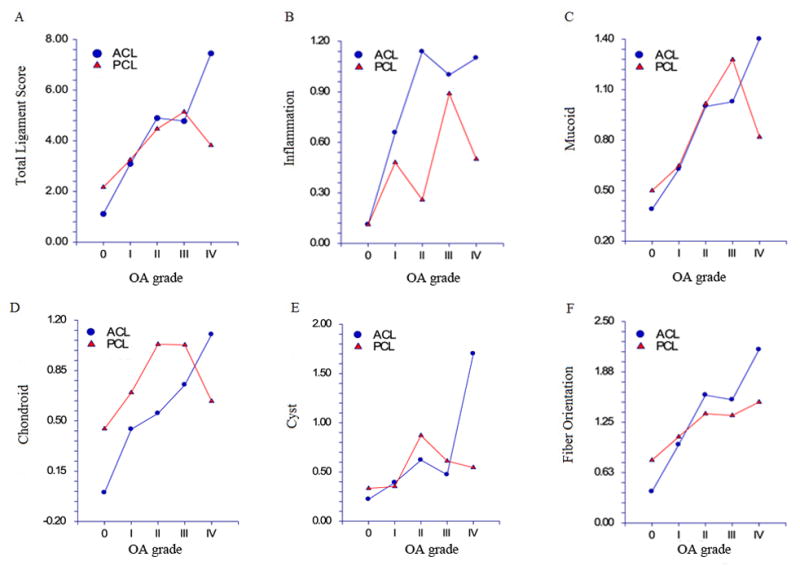
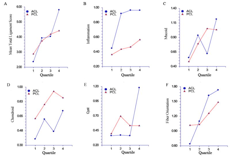
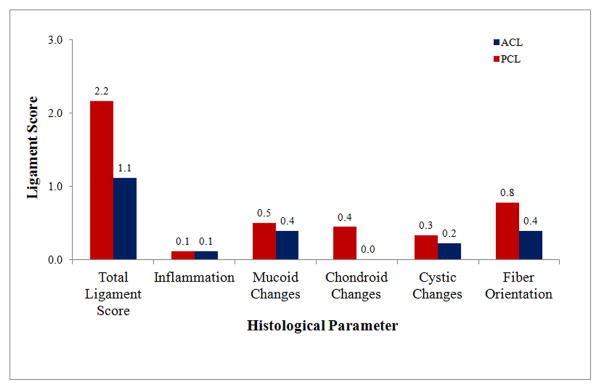
Results: All knees had intact PCL. Histologically, 6% were normal, 76% showed mild, 12% moderate and 9% severe degeneration. Fibre disorientation was the most prevalent and severe change. Histological grades of PCL and ACL correlated, but significantly fewer PCL than ACL showed severe changes. There was a weaker correlation between aging and total histological PCL scores (R=0.26) compared to aging and ACL scores (R=0.42). ACL scores correlated with cartilage scores (R=0.54) while PCL scores increased with the severity of osteoarthritis from grades 0 to III but not between osteoarthritis grades III-IV (R=0.32). In knees with ruptured ACL, the PCL scores correlated with cartilage scores of the lateral compartment.
Conclusions: PCL histopathological changes were less severe than in the ACL. PCL degeneration was associated with ACL and cartilage damage. The lack of correlation with age indicates independent pathways for PCL versus ACL degeneration.
Figures
Similar articles
-
Anterior cruciate ligament changes in the human knee joint in aging and osteoarthritis.Arthritis Rheum. 2012 Mar;64(3):696-704. doi: 10.1002/art.33417. Arthritis Rheum. 2012. PMID: 22006159 Free PMC article.
-
Predictors of posterior cruciate ligament degeneration in osteoarthritic knees.J Orthop Surg (Hong Kong). 2013 Apr;21(1):15-8. doi: 10.1177/230949901302100106. J Orthop Surg (Hong Kong). 2013. PMID: 23629980
-
Role of vascular channels as a novel mechanism for subchondral bone damage at cruciate ligament entheses in osteoarthritis and inflammatory arthritis.Ann Rheum Dis. 2015 Jan;74(1):196-203. doi: 10.1136/annrheumdis-2013-203972. Epub 2013 Oct 4. Ann Rheum Dis. 2015. PMID: 24095939 Free PMC article.
-
Histopathological analysis of the posterior cruciate ligament in primary osteoarthritis.Eur J Orthop Surg Traumatol. 2018 May;28(4):691-699. doi: 10.1007/s00590-018-2136-8. Epub 2018 Feb 7. Eur J Orthop Surg Traumatol. 2018. PMID: 29417349
-
Intraarticular Ligament Degeneration Is Interrelated with Cartilage and Bone Destruction in Osteoarthritis.Cells. 2019 Aug 27;8(9):990. doi: 10.3390/cells8090990. Cells. 2019. PMID: 31462003 Free PMC article. Review.
Cited by
-
The association between anterior cruciate ligament degeneration and incident knee osteoarthritis: Data from the osteoarthritis initiative.J Orthop Translat. 2023 Dec 14;44:1-8. doi: 10.1016/j.jot.2023.09.005. eCollection 2024 Jan. J Orthop Translat. 2023. PMID: 38174315 Free PMC article.
-
Association between magnetic resonance imaging characteristics and pathological findings in entire posterior cruciate ligament with mucoid degeneration.J Int Med Res. 2022 Mar;50(3):3000605221084865. doi: 10.1177/03000605221084865. J Int Med Res. 2022. PMID: 35272510 Free PMC article.
-
Quantitative assessment of anterior talofibular ligament quality in chronic lateral ankle instability using magnetic resonance imaging T2* value.Skeletal Radiol. 2024 Apr;53(4):733-739. doi: 10.1007/s00256-023-04480-8. Epub 2023 Oct 20. Skeletal Radiol. 2024. PMID: 37857750
-
Relationship Between Mechanoreceptors in the Posterior Cruciate Ligament and Patient Age or Osteoarthritis Severity.Orthop J Sports Med. 2023 Jun 9;11(6):23259671231168894. doi: 10.1177/23259671231168894. eCollection 2023 Jun. Orthop J Sports Med. 2023. PMID: 37332534 Free PMC article.
-
Gross Appearances of the Posterior Cruciate Ligament Correlate With Its Histological Features but not With In Vivo Function in Cruciate-Retaining Total Knee Arthroplasty.Cureus. 2023 Apr 25;15(4):e38128. doi: 10.7759/cureus.38128. eCollection 2023 Apr. Cureus. 2023. PMID: 37252485 Free PMC article.
References
-
- Felson DT, Lawrence RC, Dieppe PA, et al. Osteoarthritis: new insights. Part 1: the disease and its risk factors. Ann Intern Med. 2000;133:635–46. - PubMed
-
- Bennett LD, Buckland-Wright JC. Meniscal and articular cartilage changes in knee osteoarthritis: a cross-sectional double-contrast macroradiographic study. Rheumatology (Oxford) 2002;41:917–23. - PubMed
-
- Hill CL, Seo GS, Gale D, et al. Cruciate ligament integrity in osteoarthritis of the knee. Arthritis Rheum. 2005;52:794–9. - PubMed
-
- Bergin D, Keogh C, O’Connell M, et al. Atraumatic medial collateral ligament oedema in medial compartment knee osteoarthritis. Skeletal Radiol. 2002;31:14–8. - PubMed
Publication types
MeSH terms
Grants and funding
LinkOut - more resources
Full Text Sources
Medical
