. 2012 Sep;139(17):3180-8.
doi: 10.1242/dev.084814.

Hox genes regulate the onset of Tbx5 expression in the forelimb

Affiliations

Hox genes regulate the onset of Tbx5 expression in the forelimb

Carolina Minguillon et al. Development. 2012 Sep.

Abstract

Tbx4 and Tbx5 are two closely related T-box genes that encode transcription factors expressed in the prospective hindlimb and forelimb territories, respectively, of all jawed vertebrates. Despite their striking limb type-restricted expression pattern, we have shown that these genes do not participate in the acquisition of limb type-specific morphologies. Instead, Tbx4 and Tbx5 play similar roles in the initiation of hindlimb and forelimb outgrowth, respectively. We hypothesized that different combinations of Hox proteins expressed in different rostral and caudal domains of the lateral plate mesoderm, where limb induction occurs, might be involved in regulating the limb type-restricted expression of Tbx4 and Tbx5 and in the later determination of limb type-specific morphologies. Here, we identify the minimal regulatory element sufficient for the earliest forelimb-restricted expression of the mouse Tbx5 gene and show that this sequence is Hox responsive. Our results support a mechanism in which Hox genes act upstream of Tbx5 to control the axial position of forelimb formation.


Figures
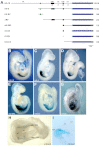
Fig. 1.
Fig. 1.
Localisation of the forelimb enhancer of the mouse Tbx5 gene. (A) Tbx5 transgenic constructs. Putative regulatory sequences (thin lines) were cloned into the BGZA reporter vector, which contains a chick β-globin minimal promoter (light-blue box), the lacZ gene (dark-blue box) and an SV40 polyadenylation signal (grey box). Black boxes represent exons (e1, e2) and the green box represents a conserved non-coding element (CNE) shared between amniotes. i1, intron 1; i2, intron 2. The numbers denote size in kb. To the right is shown the number of embryos showing forelimb-restricted expression out of the total number of transgenic embryos recovered. (B-F,H,I) Representative β-galactosidase staining for the m5-10 (B), m5-5 (C), ΔRV (D), m5-5#3 (E) and intron2 (F,H,I) constructs. (G) E9.5 mouse embryo showing endogenous Tbx5 expression assayed by whole-mount in situ hybridisation. (I) Transverse section at the forelimb level of an E9.5 intron2 transgenic embryo showing β-galactosidase staining in LPM-derived limb mesenchyme. Lateral views from the right side are shown for E9.5 embryos (B-G), whereas a ventral view with anterior to the left is shown for the E8.5 embryo (H). AER, apical ectodermal ridge; lm, limb mesoderm; LPM, lateral plate mesoderm; NT, neural tube; so, somites.
Fig. 2.
Fig. 2.
Hox4 and Hox5 paralogous group (PG) genes are expressed in the presumptive forelimb region of the LPM. (A-G) Tbx5 is expressed in the LPM of the E8 (7- to 9-somite) mouse embryo (A). PG4 and PG5 Hox genes are co-expressed in this domain: Hoxa4 (B), Hoxb4 (C), Hoxc4 (D), Hoxa5 (E), Hoxb5 (F) and Hoxc5 (G). The arrowhead marks the level of somite 3 in each embryo. (H) Fifteen-somite embryo showing endogenous expression of Tbx5. (I-N) Expression of Hoxa4 (I), Hoxb4 (J), Hoxc4 (K) and Hoxa5 (L) at the 14-somite stage and of Hoxb5 (M) and Hoxc5 (N) at the 13-somite stage. Embryos in A-G are dorsal views, anterior to the top, whereas H-N are lateral views of the right side with anterior to the top.
Fig. 3.
Fig. 3.
Hox genes can activate m5-5-driven lacZ expression in a chick co-electroporation assay. (A-F′) Co-expression of mouse (m) Hoxa4, Hoxa5, Hoxb4, Hoxb5, Hoxc4 and Hoxc5 pCIG expression plasmids (identified by expression of GFP, green in A-F) with the m5-5 BGZA reporter vector in chick hindbrain results in activation of the lacZ reporter gene (A′-F′, blue). (G) The number of β-galactosidase-positive embryos among the total number of GFP-positive embryos as a reflection of lacZ activation. This activation does not occur with the expression plasmid alone (pCIG) and is homeodomain dependent (Hoxc5 delHD). o.v., otic vesicle.
Fig. 4.
Fig. 4.
Analysis of Hox/Pbx/Meis binding sites in the intron 2 regulatory region of Tbx5. (A) Tbx5 reporter constructs in the BGZA vector used in mouse transgenesis. Blue squares, putative Hox binding sites; green squares, Hox/Meis binding sites; red squares, Pbx/Meis binding sites; yellow squares, Hox/Pbx binding sites; and grey square, a putative Tbx5 binding site. To the right is shown the number of embryos showing forelimb expression out of the total number of transgenic embryos recovered. (B-D) Dorsal views of E9.5 forelimb regions showing representative β-galactosidase staining for the intron2 MscI (B), MscI(b) (C) and Hbs3-6 (Hox3-6) (D) constructs.
Fig. 5.
Fig. 5.
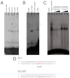
Direct binding of Hox proteins to the in-silico-predicted Hox binding sites. (A) Binding of in vitro translated mouse Hox proteins to the predicted Hox binding site 5 (Hbs5). Hoxc5 delHD provides a control. (B) Binding assay to mutant oligonucleotides and supershift assays for the predicted Hbs5. Lane 1, lysate control; lane 2, flag-tagged mouse Hoxc4 binding to labelled wild-type double-stranded oligonucleotide; lane 3, flag-tagged Hoxc4 fails to bind to labelled mutant double-stranded oligonucleotide; lane 4, supershift of the Hoxc4-Hbs5 complex with anti-flag antibody (asterisk). (C) Nuclear extracts obtained from prospective E9 forelimb regions were subjected to EMSA with labelled double-stranded oligonucleotide containing Hbs5. To test for the specificity of the shifted bands we performed competition analyses with excess unlabelled wild-type or mutant double-stranded oligonucleotide (100× molar excess, lanes 3, 6; 200×, lanes 4, 7; 500×, lanes 5, 8). (D) Double-stranded oligonucleotides used for EMSA contained mutated (mut Hox5, bold text) or wild-type (WT 5) Hbs5 (red).
Fig. 6.
Fig. 6.
Model for the activation of Tbx5 in the LPM. Hox genes belonging to PG4 and PG5 are expressed in the presumptive forelimb LPM where they bind to the intron 2 early forelimb enhancer of the mouse Tbx5 gene to specifically activate its transcription and allow Tbx5-mediated initiation of forelimb outgrowth. ex1, exon1; ex2, exon2; LPM, lateral plate mesoderm; NT, neural tube; som, somites.

Similar articles

Cited by

References

    1. Agarwal P., Wylie J. N., Galceran J., Arkhitko O., Li C., Deng C., Grosschedl R., Bruneau B. G. (2003). Tbx5 is essential for forelimb bud initiation following patterning of the limb field in the mouse embryo. Development 130, 623–633 - PubMed
    1. Boffelli D., Nobrega M. A., Rubin E. M. (2004). Comparative genomics at the vertebrate extremes. Nat. Rev. Genet. 5, 456–465 - PubMed
    1. Boulet A. M., Capecchi M. R. (1996). Targeted disruption of hoxc-4 causes esophageal defects and vertebral transformations. Dev. Biol. 177, 232–249 - PubMed
    1. Bray N., Dubchak I., Pachter L. (2003). AVID: A global alignment program. Genome Res. 13, 97–102 - PMC - PubMed
    1. Capellini T. D., Di Giacomo G., Salsi V., Brendolan A., Ferretti E., Srivastava D., Zappavigna V., Selleri L. (2006). Pbx1/Pbx2 requirement for distal limb patterning is mediated by the hierarchical control of Hox gene spatial distribution and Shh expression. Development 133, 2263–2273 - PubMed

Publication types