Review
. 2012 Nov;43(5):1873-86.
doi: 10.1007/s00726-012-1364-1. Epub 2012 Aug 8.

D-Aspartate acts as a signaling molecule in nervous and neuroendocrine systems

Affiliations
Review

D-Aspartate acts as a signaling molecule in nervous and neuroendocrine systems

Nobutoshi Ota et al. Amino Acids. 2012 Nov.

Abstract

D-Aspartate (D-Asp) is an endogenous amino acid in the central nervous and reproductive systems of vertebrates and invertebrates. High concentrations of D-Asp are found in distinct anatomical locations, suggesting that it has specific physiological roles in animals. Many of the characteristics of D-Asp have been documented, including its tissue and cellular distribution, formation and degradation, as well as the responses elicited by D-Asp application. D-Asp performs important roles related to nervous system development and hormone regulation; in addition, it appears to act as a cell-to-cell signaling molecule. Recent studies have shown that D-Asp fulfills many, if not all, of the definitions of a classical neurotransmitter-that the molecule's biosynthesis, degradation, uptake, and release take place within the presynaptic neuron, and that it triggers a response in the postsynaptic neuron after its release. Accumulating evidence suggests that these criteria are met by a heterogeneous distribution of enzymes for D-Asp's biosynthesis and degradation, an appropriate uptake mechanism, localization within synaptic vesicles, and a postsynaptic response via an ionotropic receptor. Although D-Asp receptors remain to be characterized, the postsynaptic response of D-Asp has been studied and several L-glutamate receptors are known to respond to D-Asp. In this review, we discuss the current status of research on D-Asp in neuronal and neuroendocrine systems, and highlight results that support D-Asp's role as a signaling molecule.


Figures

Fig. 1
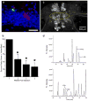
Fig. 1. D-Asp has been characterized via multiple measurement approaches
(a) Immunoreactivity measurement via a D-Asp antibody provides cellular localization, with the immunoreactivity against D-Asp observed in the anterior lobe (AL) and posterior lobe (PL), but not in the intermediate lobe (IL) in 6-week-old rat brain. Scale bar, 140 μm. (b) Chiral HPLC has been used for D-Asp characterization, with the D-Asp identified via its removal via D-aspartate oxidase (DAspO) digestion from Aplysia limacine cerebral ganglia neurons. Separation condition; C-18 column (0.45 cm×25 cm),1.2 mL/min flow rate with a programmed gradient consisting of solution A (5% acetonitrile in 30 mM citrate/phosphate buffer, pH 5.6) and solution B (90% acetonitrile in water). (c) Chiral capillary electrophoresis with nanoliter volume assays enables subcellular analysis, in this case from an individual Aplysia sensory neuron. *, unidentified peaks. Separation condition: 21 kV normal polarity was applied to an uncoated fused-silica capillary (65–75 cm, 50 μm i.d./360 μm o.d.) filled with separation solution consisting of 20 mM β-cyclodextrin, 50 mM sodium dodecyl sulfate in 50 mM borate buffer (pH 9.4) and 15% methanol (V/V). Panel a from (Lee et al. 1999), used with permission from Elsevier; panel b from (Spinelli et al. 2006) is adapted with permission, copyright © 2006 from John Wiley and Sons; and panel c from (Miao et al. 2005) is used with permission of the American Chemical Society, copyright 2005.
Fig. 2
Fig. 2. Asp racemase distribution and its tropic effect
Mouse Asp racemase was expressed in adult hippocampus and (a) the racemase expression was locally suppressed by short-hairpin RNA (shRNA) against the racemase (shRNA-DR) in newborn neurons (green fluorescence) while control shRNA did not suppress the racemase expression in newborn neurons (red fluorescence); hippocampus cells (blue) were stained with 4′,6-diamidino-2-phenylindole. Scale bar, 50 μm. (b) Survival rate of newborn neurons is compared with shRNA-DR treatment vs. shRNA-control treatment. Neurons contain both shRNA-DR and control shRNA expressed yellow fluorescence. *, P <.05; n = 4 mice for each time point, ANOVA. (c) Immunohistochemical measurement of an Asp racemase in the cerebral ganglion from Aplysia capable of racemizing both D-Asp and D-Ser. The C- and F-cluster neurons are indicated by the area enclosed in the dotted line. (d) CE analysis on C- and F-cluster neurons showed high amounts of D-Asp and D-Ser. Separation condition: 27 kV normal polarity was applied to an uncoated fused-silica capillary (80 cm total length, 70 cm effective length, 75 μm i.d./360 μm o.d.) filled with separation buffer. The separation buffer for Asp enantiomers consisted of 40 mM β-cyclodextrin and 60 mM sodium deoxycholate in 200 mM borate buffer, pH 9.5. The Ser enantiomer separation buffer consisted of 10 mM γ-cyclodextrin and 50 mM sodium dodecyl sulfate in 75 mM borate buffer, pH 10.5. Panels a and b from (Kim et al. 2010) are used with permission, copyright 2010 National Academy of Sciences, U.S.A.; panels c and d from (Wang et al. 2011) are used with permission, © the American Society for Biochemistry and Molecular Biology.
Fig. 3
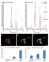
Fig. 3. D-Asp release and co-localization within synaptic vesicles
Extracellular D-Asp concentrations (a) before and (b) after potassium ion stimulation that induced D-Asp release from cerebral ganglia of Aplysia californica. In (a) and (b), the black trace (original) is the electropherogram of the original sample and the red trace (spiked) is the electropherogram of the sample spiked with 1 μM D-Asp, arrows indicate the increase in D-Asp signal due to the addition of 1 μM of standard D-Asp and the change due to potassium ion stimulation (labeled standard and stimulation, respectively). Co-localization of D-Asp and secretory granules in PC12 cells under immunofluorescent microscopic observation of (c) chromogranin A, a marker of secretory granules and (d) D-Asp; (e) merged images of c and d. Scale bar, 10 μm. Higher D-Asp content was observed in synaptic vesicles than in other cell regions in (f) rat Rattus norvegicus and (g) squid Loligo vulgaris. Panels a and b from (Scanlan et al. 2010) are used with permission, c 2010 by John Wiley & Sons, Inc.; panels c–e from (Nakatsuka et al. 2001) are used with permission, © the American Society for Biochemistry and Molecular Biology; and panels f and g are based on the data table in (D’Aniello et al. 2011).
Fig. 4
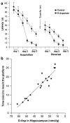
Fig. 4. Improved spatial learning and memory in rats treated with D-Asp
(a) 12 rats treated with 40 mM sodium D-Asp and 12 rats treated with 40 mM NaCl (control) were trained in the Morris water maze to measure latency in reaching the platform hidden in the maze. After the acquisition phase, the platform position was changed before starting the reversal phase, while the other settings were the same. (b) Relationship between the endogenous D-Asp in 120-day old rat hippocampus and the time to reach the platform in the Morris water maze system of the corresponding rats that were trained in the Morris water maze system. Figure is adapted from (Topo et al. 2010) and used with permission, Springer Science+Business Media.

Similar articles

Cited by

References

    1. Abe K, Takahashi S, Muroki Y, Kera Y, Yamada RH. Cloning and expression of the pyridoxal 5′-phosphate-dependent aspartate racemase gene from the bivalve mollusk Scapharca broughtonii and characterization of the recombinant enzyme. J Biochem. 2006;139(2):235–244. doi: 10.1093/jb/mvj028. - DOI - PubMed
    1. Adachi M, Koyama H, Long ZQ, Sekine M, Furuchi T, Imai K, Nimura N, Shimamoto K, Nakajima T, Homma H. L-glutamate in the extracellular space regulates endogenous D-aspartate homeostasis in rat pheochromocytoma MPT1 cells. Arch Biochem Biophys. 2004;424(1):89–96. doi: 10.1016/j.abb.2004.01.016. - DOI - PubMed
    1. Assisi L, Botte V, D’Aniello A, Di Fiore MM. Enhancement of aromatase activity by D-aspartic acid in the ovary of the lizard Podarcis s. sicula. Reproduction. 2001;121(5):803–808. doi: 10.1530/rep.0.1210803. - DOI - PubMed
    1. Bharathi, Rao KSJ, Stein R. First evidence on induced topological changes in supercoiled DNA by an aluminium D-aspartate complex. J Biol Inorg Chem. 2003;8(8):823–830. doi: 10.1007/s00775-003-0484-1. - DOI - PubMed
    1. Brown ER, Piscopo S, Chun JT, Francone M, Mirabile I, D’Aniello A. Modulation of an AMPA-like glutamate receptor (SqGluR) gating by L- and D-aspartic acids. Amino Acids. 2007;32(1):53–57. doi: 10.1007/s00726-006-0349-3. - DOI - PubMed

Publication types

MeSH terms