. 2012 Aug 29;31(17):3620-34.
doi: 10.1038/emboj.2012.224. Epub 2012 Aug 7.

The Mitotic Exit Network and Cdc14 phosphatase initiate cytokinesis by counteracting CDK phosphorylations and blocking polarised growth

Affiliations

The Mitotic Exit Network and Cdc14 phosphatase initiate cytokinesis by counteracting CDK phosphorylations and blocking polarised growth

Alberto Sanchez-Diaz et al. EMBO J. .

Abstract

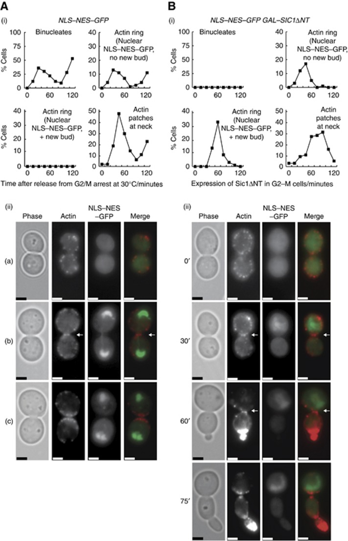
Polarisation of the actin cytoskeleton must cease during cytokinesis, to support efficient assembly and contraction of the actomyosin ring at the site of cell division, but the underlying mechanisms are still understood poorly in most species. In budding yeast, the Mitotic Exit Network (MEN) releases Cdc14 phosphatase from the nucleolus during anaphase, leading to the inactivation of mitotic forms of cyclin-dependent kinase (CDK) and the onset of septation, before G1-CDK can be reactivated and drive re-polarisation of the actin cytoskeleton to a new bud. Here, we show that premature inactivation of mitotic CDK, before release of Cdc14, allows G1-CDK to divert the actin cytoskeleton away from the actomyosin ring to a new site of polarised growth, thereby delaying progression through cytokinesis. Our data indicate that cells normally avoid this problem via the MEN-dependent release of Cdc14, which counteracts all classes of CDK-mediated phosphorylations during cytokinesis and blocks polarised growth. The dephosphorylation of CDK targets is therefore central to the mechanism by which the MEN and Cdc14 initiate cytokinesis and block polarised growth during late mitosis.


Conflict of interest statement

The authors declare that they have no conflict of interest.

Figures

Figure 1
Figure 1
Inactivation of Clb-CDK before anaphase drives cytokinesis and septation without cell separation. (A) (i) Control (W303-1a) and GAL–SIC1ΔNT (YLD12) strains were grown in YPRaff medium at 30°C and synchronised in G2–M-phase with nocodazole. Cells were switched to YPGal medium containing nocodazole for the indicated times, and cell extracts were used for immunoblots showing induction of Sic1ΔNT and dephosphorylation of the Clb-CDK target Pol12 (the loading control was the Mcm3 protein). (ii) An identical experiment was performed with 3GFP–RAS2 (YAD204) and GAL–SIC1ΔNT 3GFP–RAS2 (YASD1759). Live cells were examined by fluorescence microscopy to monitor ingression of the plasma membrane at the budneck. A white arrow denotes undivided cytoplasm at the budneck, and red arrows correspond to cells where examination of each z-level at the budneck showed divided cytoplasm. The scale bars correspond to 2 μm. (B) GAL–SIC1ΔNT ADH-OsTIR1 3GFP–RAS2 (YASD1952) and myo1-aid GAL–SIC1ΔNT ADH-OsTIR1 3GFP–RAS2 (YASD1950) were arrested with mating pheromone at 30°C in YPRaff medium, before addition of auxin and incubation for a further 60′ to deplete Myo1-aid. Cells were then released from G1-arrest and either used to monitor DNA content by flow cytometry (i), or else were synchronised in G2–M-phase with nocodazole before transfer to YPGal medium containing nocodazole and auxin (ii). Images of representative cells are shown from the 120′ time point; scale bars=2 μm (iii). (C) Control and GAL–SIC1ΔNT strains were grown as in (A), and samples were processed for electron microscopy following 120′ incubation in YPGal medium+nocodazole. The figure shows 40 nm serial sections that start just above the budneck of one control cell (nine consecutive sections) and one cell expressing GAL–SIC1ΔNT (15 consecutive sections). Additional examples are shown in Supplementary Figure 4. The scale bars correspond to 1 μm, and ‘n’ indicates the position of the nucleus in each cell.
Figure 2
Figure 2
Contraction of the actomyosin ring is defective when Clb-CDK is inactivated before anaphase. (A) Cells expressing NLS–NES–GFP and Myo1-Tomato (YASD1614) were released from nocodazole arrest at 30°C and the indicated parameters were quantified. Images (i) to (vi) show representative cells at progressively later stages of cell division and entry into the next cell cycle (NLS–NES–GFP enters both daughter nuclei at the end of anaphase upon inactivation of mitotic CDK). Scale bars=2 μm. (B) GAL–SIC1ΔNT NLS–NES–GFP MYO1-TOMATO (YASD1612) was arrested with nocodazole at 30°C as above and then switched to medium containing galactose. Representative cells are shown at the indicated time points. (C) GAL–SIC1ΔNT 3GFP–RAS2 MYO1-TOMATO (YASD1870) were treated as in (B) and images taken after 120′. Red arrows denote cells that retain a partially contracted Myo1 ring at the original budneck despite having divided their cytoplasm.
Figure 3
Figure 3
Re-polarisation of the actin cytoskeleton to a new bud competes with contraction of the actomyosin ring when Clb-CDK is inactivated before anaphase. (A) Cells expressing NLS–NES–GFP (YASD1616) were released from nocodazole arrest at 30°C as above. (i) Samples were fixed at the indicated times before staining with rhodamine-phalloidin to visualise the actin cytoskeleton. (ii) Examples are shown of a cell before assembly of the actin ring (cell (a), 0′), a cell with an actin ring (cell (b), 30′; the actin ring is denoted by a white arrow), and a cell in which the actin ring has disassembled and been replaced with patches of actin at the budneck (cell (c), 45′). (B) NLS–NES–GFP (YASD1616) and GAL–SIC1ΔNT NLS–NES–GFP (YMP197) were grown as in Figure 1A to inactivate Clb-CDK in G2–M-phase, and the cells were then fixed and stained with rhodamine-phalloidin. (i) Quantification of the indicated parameters. (ii) Examples are shown from the indicated time points; scale bars=2 μm.
Figure 4
Figure 4
Inhibition of Cln-CDK prevents re-polarisation of the actin cytoskeleton from interfering with assembly of the actin ring, when Clb-CDK is inactivated before anaphase. (A) NLS–NES–GFP MYO1-TOMATO (YASD1614) and GAL–SIC1ΔNT NLS–NES–GFP MYO1-TOMATO (YASD1612) were arrested with nocodazole at 30°C. Each culture was then split in two and transferred to medium containing galactose plus nocodazole, with or without α-factor mating pheromone. (B) A similar experiment was performed with NLS–NES–GFP (YASD1616) and GAL–SIC1ΔNT NLS–NES–GFP (YMP197) and samples were fixed and stained with rhodamine-phalloidin as above. (i) For each sample, 100 cells were examined to determine the percentage in which the actin cytoskeleton was polarised to the site of a new bud (−α-factor) or a shmoo (+α-factor). (ii) Examples of post-cytokinesis cells with shmoos; scale bars=2 μm. (iii) The percentage of cells with actin rings was determined for the same experiment.
Figure 5
Figure 5
Inhibition of Clb-CDK without reactivation of Cln-CDK induces contraction of the actomyosin ring and the rapid completion of cytokinesis. (A) The proportion of cells with Myo1 rings at the budneck was determined for the experiment in Figure 4A. (B) A similar experiment was performed with GAL–SIC1ΔNT 3GFP–RAS2 (YASD1759), to monitor completion of cytokinesis. (C) The experiment in Figure 4A was repeated and time-lapse video microscopy was used to monitor the relation between Clb-CDK inactivation and contraction of the actomyosin ring. Note that re-budding was inhibited in this experiment by illumination of the field of view with UV light. The times at which contraction of the Myo1 ring began and was completed are indicated by white and black circles, respectively.
Figure 6
Figure 6
An increase in the pool of free Cdc14 improves the efficiency of cytokinesis and actomyosin-ring contraction, when Clb-CDK is inactivated before anaphase. (A) GAL–SIC1ΔNT 3GFP–RAS2 (YASD2047), GAL–CDC14 3GFP–RAS2 (YASD2049) and GAL–SIC1ΔNT GAL–CDC14 3GFP–RAS2 (YASD2041) were grown as in Figure 1A. Red arrows denote the budneck region of cells with divided cytoplasm. The scale bar corresponds to 5 μm. (B) Analogous experiments were performed with GAL–SIC1ΔNT NLS–NES–GFP MYO1-TOMATO (YASD1612), GAL–CDC14 NLS–NES–GFP MYO1-TOMATO (YASD2005) and GAL–SIC1ΔNT GAL–CDC14 NLS–NES–GFP MYO1-TOMATO (YASD2001).
Figure 7
Figure 7
Defective cytokinesis following Clb-CDK inactivation in MEN mutants is associated with defects in Cdc14 function. (A) GAL–SIC1ΔNT 3GFP–RAS2 (YASD1870) and cdc14-1 cdc15-2 GAL–SIC1ΔNT 3GFP–RAS2 (YASD1852) were arrested with nocodazole at 24°C in medium containing raffinose, before switching cells to 37°C for 60′. An aliquot of cells was released from nocodazole at this point to confirm that inactivation of Cdc14 and Cdc15 caused cells to arrest after anaphase (not shown). The remainder of the culture was switched to medium containing galactose and nocodazole, and incubation continued at 37°C for the indicated times. Scale bars=5 μm. (B) dbf2-2 dbf20Δ GAL–SIC1ΔNT 3GFP–RAS2 (YASD2154) was compared with the control strain in a similar experiment. (C) An analogous experiment to that in (A) was performed with GAL–SIC1ΔNT NLS–NES–GFP MYO1-TOMATO (YASD1612) and cdc14-1 cdc15-2 GAL–SIC1ΔNT NLS–NES–GFP MYO1-TOMATO (YASD1926). (D) dbf2-2 dbf20Δ GAL–SIC1ΔNT NLS–NES–GFP MYO1-TOMATO (YASD2111) was compared with the control strain (YASD2093) in a similar experiment.
Figure 8
Figure 8
Cytoplasmic Cdc14 largely rescues the cytokinesis defects of dbf2-2 dbf20Δ. (A) (i) GAL–SIC1ΔNT 3GFP–RAS2 (YASD2047), dbf2-2 dbf20Δ GAL–SIC1ΔNT 3GFP–RAS2 (YASD2154) and dbf2-2 dbf20Δ GAL–SIC1ΔNT GAL–cdc14-BP1,2A 3GFP–RAS2 (YASD2179) were grown and processed as in Figure 7, to monitor progression through cytokinesis. (ii) Analogous experiments were performed with dbf2-2 dbf20Δ GAL–SIC1ΔNT 3GFP–RAS2 (YASD2154), dbf2-2 dbf20Δ GAL–SIC1ΔNT GAL–CDC14 3GFP–RAS2 (CC8336) and dbf2-2 dbf20Δ GAL–SIC1ΔNT GAL–cdc14–BP1,2A 3GFP–RAS2 (YASD2179). (B) Representative images from the experiments in (A). Red arrows denote cells with divided cytoplasm at the budneck. Scale bars=2 μm.

Similar articles

Cited by

References

    1. Akiyoshi B, Biggins S (2010) Cdc14-dependent dephosphorylation of a kinetochore protein prior to anaphase in Saccharomyces cerevisiae. Genetics 186: 1487–1491 - PMC - PubMed
    1. Amon A, Tyers M, Futcher B, Nasmyth K (1993) Mechanisms that help the yeast cell cycle clock tick: G2 cyclins transcriptionally activate G2 cyclins and repress G1 cyclins. Cell 74: 993–1007 - PubMed
    1. Bardin AJ, Amon A (2001) MEN and SIN: what's the difference? Nat Rev Mol Cell Biol 2: 815–826 - PubMed
    1. Barr FA, Gruneberg U (2007) Cytokinesis: placing and making the final cut. Cell 131: 847–860 - PubMed
    1. Bembenek J, Kang J, Kurischko C, Li B, Raab JR, Belanger KD, Luca FC, Yu H (2005) Crm1-mediated nuclear export of Cdc14 is required for the completion of cytokinesis in budding yeast. Cell cycle 4: 961–971 - PubMed

Publication types

MeSH terms