Review
. 2013:757:133-70.
doi: 10.1007/978-1-4614-4015-4_6.

Meiotic development in Caenorhabditis elegans

Affiliations
Review

Meiotic development in Caenorhabditis elegans

Doris Y Lui et al. Adv Exp Med Biol. 2013.

Abstract

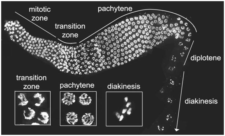
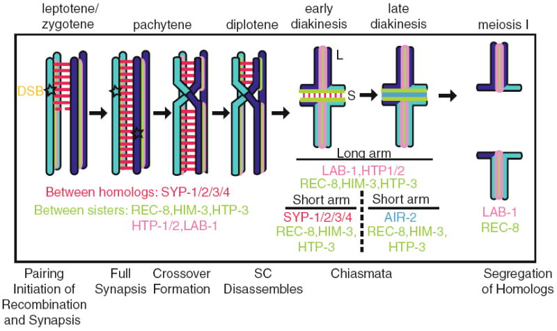
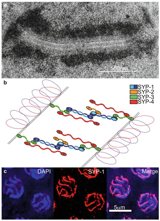
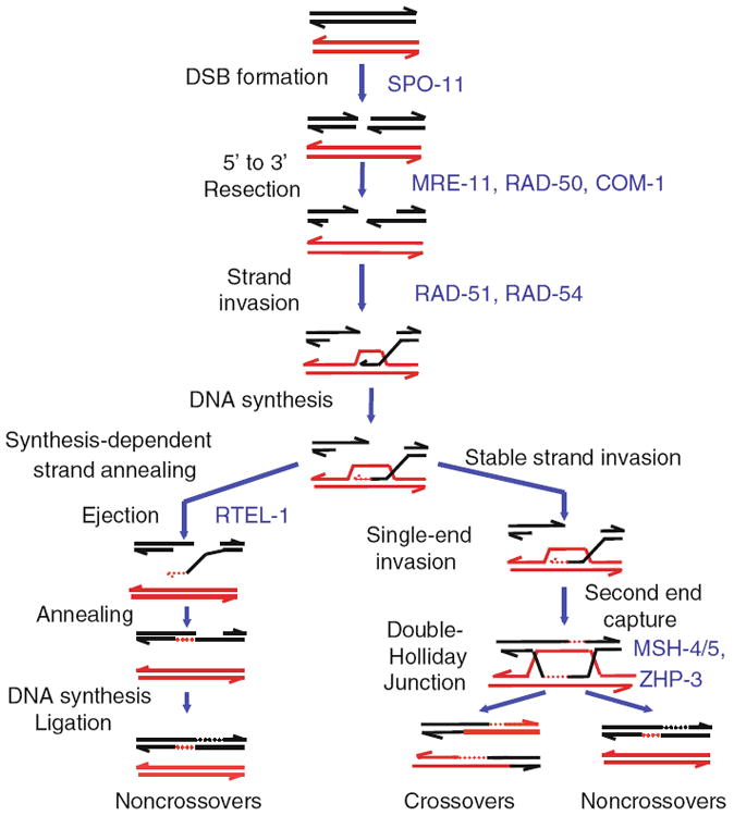
Caenorhabditis elegans has become a powerful experimental organism with which to study meiotic processes that promote the accurate segregation of chromosomes during the generation of haploid gametes. Haploid reproductive cells are produced through one round of chromosome replication followed by two -successive cell divisions. Characteristic meiotic chromosome structure and dynamics are largely conserved in C. elegans. Chromosomes adopt a meiosis-specific structure by loading cohesin proteins, assembling axial elements, and acquiring chromatin marks. Homologous chromosomes pair and form physical connections though synapsis and recombination. Synaptonemal complex and crossover formation allow for the homologs to stably associate prior to remodeling that facilitates their segregation. This chapter will cover conserved meiotic processes as well as highlight aspects of meiosis that are unique to C. elegans.


Figures

Fig. 6.1
Fig. 6.1
The C. elegans gonad. A dissected and DAPI-stained gonad of a hermaphrodite adult worm. Progression from the distal to the proximal end is depicted from left to right. The image is a projection of three-dimensional data stacks of intact nuclei, which were taken approximately halfway through the gonad, to facilitate visualization of nuclear morphology
Fig. 6.2
Fig. 6.2
Events during meiotic progression that contribute to the proper segregation of homologs during the first meiotic division
Fig. 6.3
Fig. 6.3
The synaptonemal complex. (a) TEM (transmission electron microscopy) image of the structure of the SC between chromosomes in a pachytene nucleus in the C. elegans germline. The continuous zipper-like track, comprised of the transverse filaments, is flanked by electron-dense patches of chromatin. (b) Schematic of the arrangement of the four central region proteins in the SC of C. elegans. (c) Immunolocalization of SC proteins in pachytene nuclei. Central region protein SYP-1 (red) forms tracks at the interface between DAPI-stained chromosomes (blue). The images are projections halfway through three-dimensional data stacks of whole nuclei
Fig. 6.4
Fig. 6.4
The meiotic recombination pathway in C. elegans. Here one homolog is depicted in black and the other is shown in red. Meiotic recombination is initiated by formation of DSBs. The topoisomerase-like enzyme SPO-11 catalyzes the cleavage of the double-stranded DNA of one sister chromatid. Both 5′ ends of the DSB are rapidly resected by MRE-11, RAD-50, and COM-1 to reveal 3′ single-stranded tails, on which RAD-51 forms a filament. RAD-54 promotes invasion by one end of the DSB into the DNA duplex of the homolog to form the nascent D-loop structure. As DNA synthesis occurs, the D-loop expands. The D-loop structure is processed by two major pathways to yield COs and NCOs. COs, which hold bivalents together, arise from the formation of stable single-end invasions, followed by second end capture and then the formation of double Holliday junctions, which are cleaved by a currently unknown resolvase. NCOs arise from either processing of double Holliday junctions or the synthesis-dependent strand-annealing pathway, through which the invading end of the DSB is ejected so that it can anneal with its sister chromatid. Following annealing, DNA synthesis and ligation occur to complete the formation of NCOs

References

    1. Adamo A, Montemauri P, Silva N, Ward JD, Boulton SJ, La Volpe A. BRC-1 acts in the inter-sister pathway of meiotic double-strand break repair. EMBO Rep. 2008;9(3):287–292. - PMC - PubMed
    1. Agarwal S, Roeder GS. Zip3 provides a link between recombination enzymes and synaptonemal complex proteins. Cell. 2000;102(2):245–255. - PubMed
    1. Albertson DG, Thomson JN. The kinetochores of Caenorhabditis elegans. Chromosoma. 1982;86(3):409–428. - PubMed
    1. Alexeev A, Mazin A, Kowalczykowski SC. Rad54 protein possesses chromatin-remodeling activity stimulated by the Rad51-ssDNA nucleoprotein filament. Nat Struct Biol. 2003;10(3):182–186. - PubMed
    1. Allard P, Colaiacovo MP. Bisphenol A impairs the double-strand break repair machinery in the germline and causes chromosome abnormalities. Proc Natl Acad Sci USA. 2010;107(47):20405–20410. - PMC - PubMed

Publication types

MeSH terms

LinkOut - more resources