Review
. 2013:757:171-203.
doi: 10.1007/978-1-4614-4015-4_7.

Spermatogenesis

Affiliations
Review

Spermatogenesis

Diana S Chu et al. Adv Exp Med Biol. 2013.

Abstract

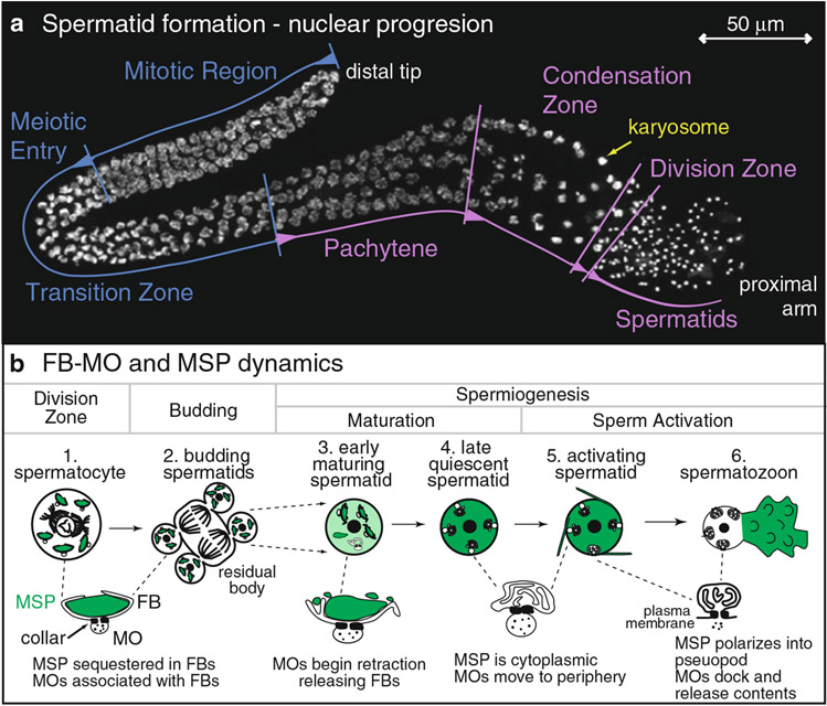
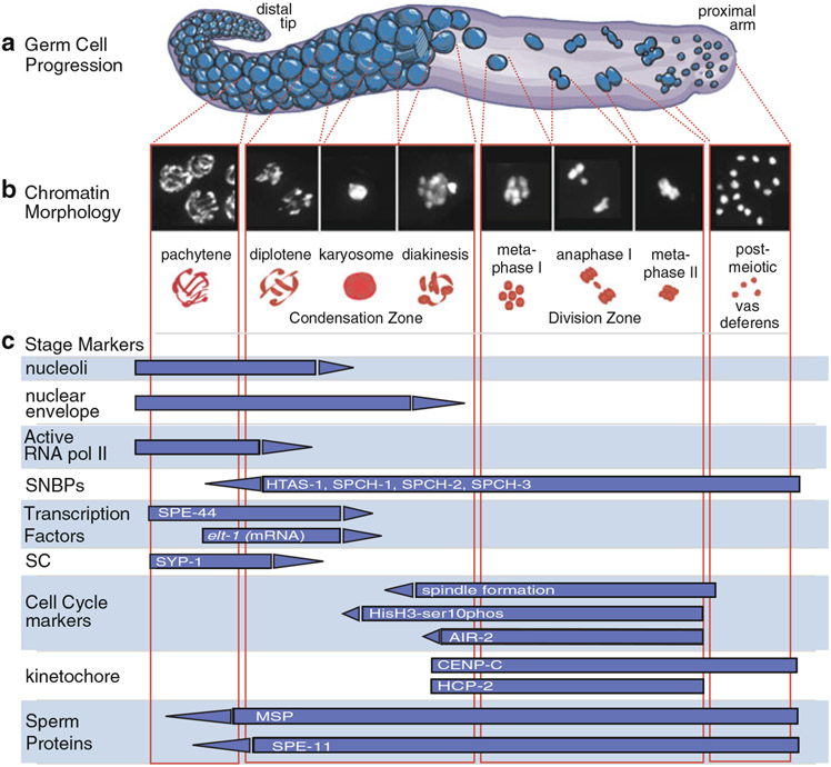
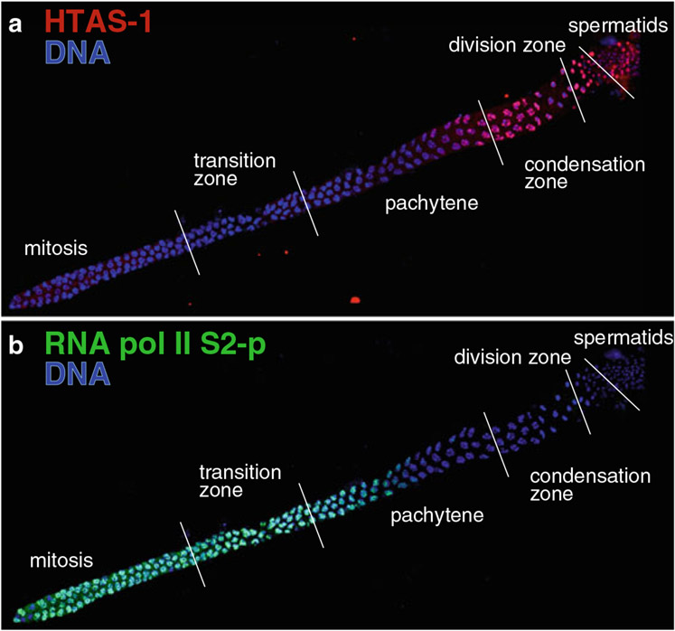
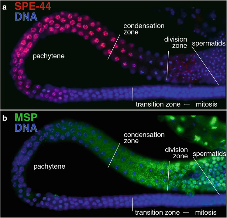
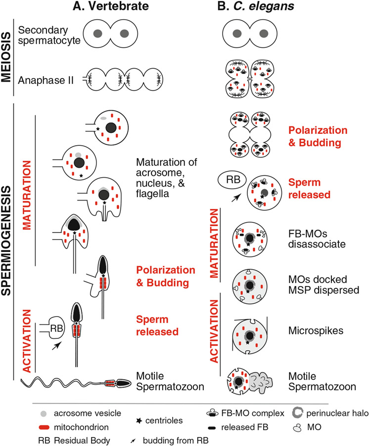
During spermatogenesis, pluripotent germ cells differentiate to become efficient delivery vehicles to the oocyte of paternal DNA. Though male and female germ cells both undergo meiosis to produce haploid complements of DNA, at the same time they also each undergo distinct differentiation processes that result in either sperm or oocytes. This review will discuss our current understanding of mechanisms of sperm formation and differentiation in Caenorhabditis elegans gained from studies that employ a combination of molecular, transcriptomic, and cell biological approaches. Many of these processes also occur during spermatogenesis in other organisms but with differences in timing, molecular machinery, and morphology. In C. elegans, sperm differentiation is implemented by varied modes of gene regulation, including the genomic organization of genes important for sperm formation, the generation of sperm-specific small RNAs, and the interplay of specific transcriptional activators. As sperm formation progresses, chromatin is -systematically remodeled to allow first for the implementation of differentiation programs, then for sperm-specific DNA packaging required for transit of paternal genetic and epigenetic information. Sperm also exhibit distinctive features of -meiotic progression, including the formation of a unique karyosome state and the centrosomal-based segregation of chromosomes during symmetric meiotic -divisions. Sperm-specific organelles are also assembled and remodeled as cells complete -meiosis and individualize in preparation for activation, morphogenesis, and the acquisition of motility. Finally, in addition to DNA, sperm contribute specific cellular factors that contribute to successful embryogenesis.


Figures

Fig. 7.1
Fig. 7.1
The progression of spermatid formation and pseudopod assembly. (a) Changes in nuclear morphology during spermatid formation. A single-armed wild-type male gonad visualized using DAPI and fluorescence microscopy. Regions of the gonad are labeled: mitotic, meiotic entry, and the transition zone (blue) and pachytene, the condensation zone, meiotic division zone, and haploid spermatids (purple). A nucleus exhibiting the karysome morphology is indicated in yellow. Scale bar represent 50 μm. (b) Key stages in FB-MO and MSP dynamics. The process of spermiogenesis includes budding, maturation, and activation. (1) A spermatocyte in diakinesis (before nuclear envelope breakdown) contains multiple, fully mature fibrous body (FB)–membranous organelle (MO) complexes. The major sperm protein (MSP) (green) is assembled into FBs that are enveloped by the arms of the MO. The MO head is the vesicle below the electron-dense collar (two dark bars). (2) After the MI and MII divisions the late-stage budding spermatid is fully polarized with the FB-MOs and chromatin masses partitioned to the spermatids and the intact spindle microtubules partitioned to the central residual body. (3) In an early maturing spermatid, the arms of the MO retract as the FBs are released into the cytoplasm and begin to disassemble. (4) A late-stage quiescent spermatid in which the MOs are docked and MSP is cytosolic. (5) Upon exposure to an activator, spermatids initially form microspikes as the MOs begin to fuse at the collar with the plasma membrane. (6) Motile spermatozoon with a distinct cell body containing fused MOs and MSP-filled pseudopod
Fig. 7.2
Fig. 7.2
Overview of the overlapping events that occur during late meiotic prophase of sperm cell formation. (a) Schematic of the progression of male germline cells (blue) during spermatogenesis. Cells are attached to the rachis through karyosome formation, then bud off of the rachis to undergo meiotic divisions. After anaphase II, haploid cells bud from residual bodies to form spermatids. (b) The corresponding chromatin morphology of cells highlighted in (a). DAPI-stained and schematic drawings (red) of the nuclear morphology of cells in the stages of late meiotic prophase indicated. (c) Staging of sperm cells can also be monitored by the presence of specific cell structures, organelles, and macromolecules, which are represented as blue bars
Fig. 7.3
Fig. 7.3
Changes in transcriptional regulation correlate with the alteration of chromatin composition. Immunolocalization of (a) the sperm-specific histone H2A variant, HTAS-1 (red) and (b) elongating RNA polymerase II (phosphorylated on the C-terminal domain on serine 2, detected using Abcam H5 antibody ab24758) (green). Regions of the male germ line are indicated. DNA is shown in blue. (a) HTAS-1 incorporates into sperm chromatin as cells condense for meiotic divisions. (b) High levels of actively elongating RNA polymerase (green) decrease dramatically as chromosomes condense for meiotic divisions, indicating global transcriptional activation is curtailed by the karyosome stage
Fig. 7.4
Fig. 7.4
Sperm-specific transcription factors and the expression of sperm proteins. Immunolocalization of (a) the SPE-44 transcription factor (red) and (b) the Major Sperm Protein, MSP (green) within an isolated and fixed male gonad. DNA is shown in blue. Regions of the male germ line are indicated. (a) SPE-44 (green) is expressed early in pachytene until chromosomes condense for meiotic divisions. (b) MSP is synthesized beginning in pachytene and subsequently localizes to distinct FBs, which localize as oblong stripes,within the condensation zone. MSP partitions to the spermatids in FBs but in mature, quiescent spermatids (far right) it disassembles and fills the cytoplasm
Fig. 7.5
Fig. 7.5
Comparison of spermiogenesis in (a) vertebrates and (b) C. elegans, highlighting analogous events. (a) In vertebrates, following anaphase II, spermatocytes undergo incomplete cytokinesis to generate four, interconnected haploid spermatids. These spermatids then undergo a multi-week maturation process of spermiogenesis that includes the following events: a burst of sperm-specific transcription and translation, the formation of a mature acrosome, the mature flagellum, and the compaction and reshaping of the nucleus. Materials unneeded by the spermatozoon are then partitioned into a residual body (RB) as the spermatozoon completes cellularization. Sperm activation causes a spermatozoon to become fully motile. (b) In C. elegans, following anaphase II, spermatocytes initiate a cleavage furrow that regresses and morphs into a polarization and budding process during which time unneeded materials are partitioned away from the differentiating sperm and left in a central residual body as spermatids detach. During a short (minutes-long) maturation step the MOs mature and dock, the FBs disassociate and subsequently disassemble, and an RNA-enriched perinuclear halo forms around the compact chromatin mass. Male spermatids are stored in this quiescent state until stimulated by extracellular signals to active and form bipolar, motile spermatozoa. Both hermaphrodite and male sperm activation occurs in less than 10 minutes
Fig. 7.6
Fig. 7.6
Signaling and response elements involved in sperm activation. (a) Diagram of a two-pathway model for sperm activation. The SWM-1 protease inhibitor is present in both males and hermaphrodites, but its only known target to date is the male-specific protease TRY-5. The action of TRY-5 as a component of a male-specific activation pathway may be direct or indirect. The cellular response pathway downstream of TRY-5 (Pathway “X”) is present in both male and hermaphrodite sperm. The SPE-8 group components comprise a second cellular response pathway, which is also present in both male and hermaphrodite sperm. The in vivo activator of this SPE-8 pathway has yet to be either molecularly or mutationally identified. This unknown activator is definitely present in hermaphrodites, and it may or may not be redundantly present in males. The in vitro activator Pronase activates sperm via the SPE-8 pathway. In vitro activation by weak bases bypasses the absence of either TRY-5 or SPE-8 group components. The two cellular response pathways converge to inhibit the SPE-6 kinase. (b) Hypothetical model for sperm activation events (adopted from Gosney et al. 2008). Close-up views of the region indicated by the gray boxes are shown on the schematic of docked or fused MOs below. In quiescent spermatids, cytosolic SPE-6 actively phosphorylates and thus inhibits the MO membrane protein SPE-4. Upon sperm activation, signaling components activate to inhibit SPE-6. As a result, SPE-4 becomes active and can cleave FER-1. The proteolytically processed form of FER-1 promotes MO fusion with the plasma membrane

References

    1. Achanzar WE, Ward S (1997) A nematode gene required for sperm vesicle fusion. J Cell Sci 110(Pt 9):1073–1081 - PubMed
    1. Aitken RJ, De Iuliis GN (2007) Origins and consequences of DNA damage in male germ cells. Reprod Biomed Online 14(6):727–733 - PubMed
    1. Albertson DG (1984) Formation of the first cleavage spindle in nematode embryos. Dev Biol 101(1):61–72 - PubMed
    1. Albertson DG, Thomson JN (1993) Segregation of holocentric chromosomes at meiosis in the nematode, Caenorhabditis elegans. Chromosome Res 1(1):15–26 - PubMed
    1. Aoki K, Moriguchi H, Yoshioka T, Okawa K, Tabara H (2007) In vitro analyses of the production and activity of secondary small interfering RNAs in C. elegans. EMBO J 26(24):5007–5019 - PMC - PubMed

Publication types

MeSH terms